RESEARCH ARTICLE
Liposomal Gel of Centella asiatica: Antioxidant Activity and Release Profile
Academic Editor: Abdelrahman I. Rezk
Sciences of Pharmacy|Vol. 4, Issue 4, pp. 328-337 (2025)
Views
Downloads
Shares
Received
Sep 9, 2025Revised
Nov 12, 2025Accepted
Nov 30, 2025Published
Dec 30, 2025
Abstract
Introduction
Wound healing is a complex and highly regulated biological process that can be severely disrupted by oxidative stress, inflammation, and impaired microcirculation. Excessive production of reactive oxygen species (ROS) is a major contributor to delayed epithelialization and the development of chronic wounds. This underscores the need for therapeutic strategies with a strong antioxidant capacity to support tissue regeneration (1). Topical drug delivery is promising for wound management because it allows for localized treatment while minimizing systemic exposure; however, achieving adequate dermal penetration remains challenging due to the barrier properties of the stratum corneum (2).
Centella asiatica (L. ) Urban is well known for its wound-healing properties, primarily due to its abundance of bioactive compounds, such as triterpenoids (including asiaticoside, madecassoside, and asiatic acid) and flavonoids, such as quercetin. These constituents work together to promote collagen synthesis, stimulate angiogenesis, and provide antioxidant protection (3). However, the topical application of C. asiatica extract is significantly hindered by its poor aqueous solubility and rapid degradation, which leads to low bioavailability. This issue is further compounded by the multilayered structure of the skin, particularly the stratum corneum, which acts as a highly lipophilic and tightly packed barrier. This barrier restricts the diffusion of hydrophilic and unstable compounds, resulting in only a minimal amount of the active ingredients penetrating deeper skin layers. Consequently, the therapeutic effectiveness of C. asiatica when applied topically is reduced (4,5).
It is important to justify the use of whole-plant extracts over isolated compounds like pure quercetin. Polyphenolic combinations found in full-spectrum extracts exhibit greater biological efficacy than isolated molecules due to their additive and synergistic effects on inflammatory and ROS-regulating pathways (6). Therefore, encapsulating whole extracts may provide therapeutic advantages that purified compounds cannot offer alone.
Advanced nanocarrier-based delivery systems, such as liposomes embedded in hydrogels, have shown improved dermal penetration, enhanced entrapment efficiency, and sustained drug release profiles for botanical phytochemicals. Recent studies investigating liposomal gels containing extracts from Vitis vinifera, Withania somnifera, and Ceratonia siliqua have reported significant improvements in wound closure and antioxidant performance (7-9). However, despite the growing scientific interest in C. asiatica, there is a limited number of studies specifically focused on optimizing liposomal hydrogel formulations that encapsulate ethanolic extracts of C. asiatica, as well as comparing the effects of extract concentration on antioxidant activity and release behavior. This represents a significant research gap.
hydrogels serve as effective vehicles, offering good spreadability, hydration, and controlled release characteristics (4,5). The use of liposomal gel systems has gained significant attention due to their ability to improve stability, enhance dermal delivery, and prolong the therapeutic effects of bioactive compounds (2,10). Based on this rationale, we hypothesize that the incorporation of C. asiatica ethanolic extract into liposomal gel formulations enhances physicochemical stability, preserves antioxidant activity, and improves transdermal penetration. Accordingly, this study aims to formulate and evaluate liposomal gels containing C. asiatica extract at two concentrations (0.3% and 0.5%) and to assess their antioxidant activity and in vitro release and permeation profiles as a basis for the development of topical wound-healing formulations.
Methodology or Experimental Section
Equipsments
The instruments used in this study included a UV–Vis spectrophotometer (UV Mini-60, Shimadzu, Japan), vacuum rotary evaporator (RV 10 Basic, IKA, Germany), centrifuge (Z 216 MK, HERMLE, Germany), particle size analyzer (SZ-100, HORIBA Scientific, Japan), probe sonicator (JY92-IIDN, Scientz, China), analytical balance (Pioneer PX Series, OHAUS, Germany/USA), laboratory oven (UN55, Memmert, Germany), refrigerator (RT Series, Panasonic, Japan), pH meter (SevenCompact F20, Mettler Toledo, Switzerland), water bath (HH-S6, Jintan, China), moisture balance (MB120, OHAUS, USA), and Franz diffusion cell apparatus (PermeGear, USA).
Materials
C. asiatica (L. ) Urban (pegagan) was collected from Bandorasa Kulon Village, Cilimus Subdistrict, Kuningan Regency, West Java, Indonesia, and authenticated by the Plant Taxonomy Laboratory, Department of Biology, FMIPA, Padjadjaran University (Plant Identification Certificate No. 54/HB/12/2023). Reagents were obtained from PT Pratama Sains Global, Indonesia (aquadest, soy lecithin, 70% ethanol, AlCl₃, sodium acetate, vitamin C, DPPH, carbopol, propylene glycol, methyl paraben, TEA), Merck KGaA, Germany (absolute ethanol, NaOH, KH₂PO₄), Sigma-Aldrich, USA (cholesterol, chloroform, methanol), and HiMedia Laboratories Pvt. Ltd. , India (quercetin standard). All materials were of analytical grade to ensure experimental reliability and reproducibility.
Methods
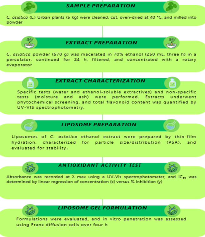
The image above shows the research methodology flow, starting from C. asiatica sample preparation, extraction, and characterization processes, to liposome gel formulation and antioxidant activity testing, as illustrated in Figure 1.
Extract characterization
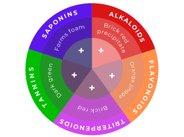
Extracts were characterized using specific parameter tests (water-soluble juice content and ethanol-soluble juice content) and non-specific parameters (water content and ash content). Phytochemical screening was conducted by analyzing the content of alkaloids, flavonoids, triterpenoids, tannins, and saponins.
The determination of total flavonoid
The total flavonoid was measured by measuring the absorbance of the sample solution using UV-Vis spectrophotometry. The standard curve for quercetin was determined using five concentration series: 20, 30, 40, 50, and 60 ppm. The absorbance was measured at a wavelength of 430.5 nm after a 30-minute operating time. The total flavonoid content was expressed as quercetin equivalents (QE), and all measurements were performed in triplicate to ensure accuracy and reproducibility.
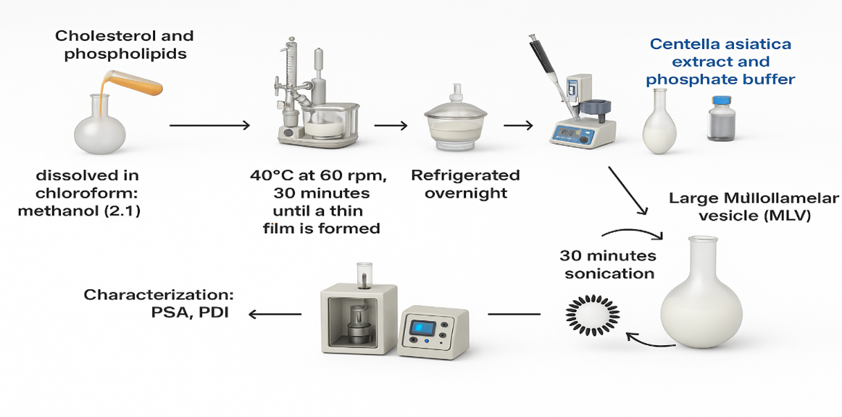
Liposome preparation using the thin-layer hydration method (11).
Liposome preparation was carried out using the thin-layer hydration method, as schematically illustrated in Figure 2. Liposomes were prepared by the thin-film hydration method with minor modifications in lipid ratio and hydration conditions. Lipid base (soy lecithin; cholesterol) was dissolved in 15 mL of chloroform: methanol (2: 1) solution in an evaporator flask (40°C at 60 rpm). Then, the formed film was left in a desiccator overnight. Then, it was hydrated with phosphate buffer (pH 7.4) containing an ethanol extract of C. asiatica (L. ) Urban, up to 20 mL in a rotary evaporator for 120 min at 60 °C and 200 rpm (12). Then, the particle size and distribution of the liposome formula of C. asiatica (L. ) Urban ethanol extracts were measured using a particle size analyzer, and a stability test was conducted. Table 1 shows the composition of the C. asiatica (L. ) Urban ethanol liposome formula.
| No. | Material | Formulation (%) | Usability | ||
|---|---|---|---|---|---|
| Basis | FG1 | FG2 | |||
| 1. | C. asiatica (L. ) Urban extract liposomes | - | 0.3 | 0.5 | Active Substance |
| 2. | Carbopol 940 | 0, 5 | 0, 5 | 0, 5 | Gelling Agent |
| 3. | Propylenglycol | 7, 5 | 7, 5 | 7, 5 | Humectants |
| 4. | Methylparaben | 0, 2 | 0, 2 | 0, 2 | Preservatives |
| 5. | Triethanolamine (TEA) | 0, 14 | 0, 14 | 0, 14 | Alkalizing Agent |
| 6. | Aquadest (mL) | add 100 | add 100 | add 100 | Solvent |
Antioxidant Activity Test
Antioxidant activity was evaluated using the DPPH radical scavenging assay with vitamin C as the positive control (13,14). The preparation of a 100 ppm DPPH solution was then followed by the determination of the maximum wavelength and operating time. The maximum wavelength was determined using 4 mL of methanol and 3 mL of DPPH solution. The absorbance was then measured at wavelengths of 400-800 nm, and the operating time was determined at 2 min intervals. Preparation of a standard solution of vitamin C 100 ppm, standard solution of C. asiatica (L. ) Urban extract 100 ppm, standard solution of liposome formula I 100 ppm, and standard solution of liposome formula II 100 ppm. Each standard solution was diluted into a master solution with concentrations of 4 ppm, 6 ppm, 8 ppm, and 10 ppm. The sample was then incubated in a brown vial at room temperature during the operating time. Absorbance was recorded at 515 nm after 30 min of reaction. Methanol without DPPH served as the blank, and vitamin C as the positive control. A standard calibration curve was generated, and IC₅₀ was calculated from triplicate experiments (n = 3). The IC50 value of each sample was determined.
The resulting absorbance value is then entered into the % inhibition formula. A standard curve was then created between concentration (ppm) and % inhibition. The IC50 value was calculated using a linear regression equation of y bx + a between the concentration of the test solution (x) and the %inhibition (y).
Liposome Gel Formulation Preparation of ethanol extract of C. asiatica (L. ) Urban
Liposome gel containing ethanol extract of C. asiatica (L. ) Urban was prepared using Carbopol gel base. 0.5% Carbopol was accurately weighed and dispersed in distilled water, and then it was kept for 30 min. Then, triethanolamine and methylparaben dissolved in propylene glycol were added to the gel base, and liposomes of C. asiatica (L. ) Urban ethanol extract was added to the FG1 and FG2 formulations. Both gels were homogenized using a homogenizer with a stirring speed of 500 rpm for 15 min, and their properties were evaluated in terms of organoleptic characteristics, homogeneity, spreadability, pH, viscosity, and rheology. Details of the liposome gel formulation of ethanol extract of C. asiatica (L. ) Urban is shown in Table 2.
| Material | Formula | Usability | ||
|---|---|---|---|---|
| F0 | F1 | F2 | ||
| Ethanol Extract of C. asiatica (L. ) Urban | - | 0.3 % | 0.5 % | Active Substance |
| Soya Lecithin | 90 mg | 90 mg | 90 mg | Liposome constituents |
| Cholesterol | 10 mg | 10 mg | 10 mg | Liposome constituents |
| phosphate buffer pH 7.4 | 20 mL | 20 mL | 20 mL | Buffer |
In-Vitro Penetration Test
In vitro skin permeation was performed using static Franz diffusion cells with minor adaptations in sampling schedule and receptor composition (15). Skin permeation was evaluated using Franz diffusion cells, which have a receptor volume of 20 mL and a diffusion area of 2.5 cm². Wistar rat abdominal skin (Rattus norvegicus) was used for this study, following ethics approval from the Health Research Ethics Committee at Universitas Muhammadiyah Ahmad Dahlan Cirebon (Protocol No. 002/VII/2025/0004/KEPK/STFMC, valid from July 2025 to July 2026). The receptor chamber contained phosphate-buffered saline (pH 7.4) maintained at a temperature of 37 ± 0.5 °C with constant stirring. Aliquots of 1 mL were withdrawn at predetermined intervals (0–240 min) and replaced with pre-warmed medium to ensure sink conditions, in accordance with established diffusion-kinetic procedures. 1 g of gel was placed between the donor compartment and the receptor compartment with the stratum corneum facing upward. Samples were taken at 30, 60, 90, 120, 180, and 240 min intervals and analyzed using a UV-Vis spectrophotometer at 374.5 nm.
Results and Discussion
Phytochemical Screening and Characterization of the Ethanol Extract of C. asiatica (L. ) Urban
The extraction yield indicates how effectively the solvent can extract the compound from the simplisia (16). The ethanol extract of C. asiatica (L. ) Urban obtained 96.14 g of thick extract from 570 g of simplicia, yielding 16.86%. This value exceeds both the Indonesian Herbal Pharmacopoeia (FHI) requirement of not less than 7.2% and the general requirement for thick extracts, which must be at least 10% (17). Therefore, it can be concluded that the yield of the ethanol extract of C. asiatica meets the established pharmacopoeial standards and fulfills the required quality criteria.
Qualitative phytochemical screening tests were conducted as preliminary evaluations on ethanol extracts of C. asiatica(L. ) Urban et al. determined the presence of secondary metabolite content using color reagents (18). Preliminary tests conducted in this study include tests for flavonoid compounds, alkaloids, saponins, and tannins. The results of the phytochemical screening carried out show that the ethanol extract of C. asiatica(L. ) Urban contains flavonoids, triterpenoids, tannins, alkaloids, and saponins (see Figure 3). A qualitative phytochemical screening was conducted using colorimetric reagents on the ethanol extract of C. asiatica (L. ) Urban as a preliminary assessment to identify the major classes of secondary metabolites. The results of the extract profile indicated the presence of flavonoids, triterpenoids/terpenoids, tannins, and saponins, as well as, in some reports, alkaloids. The variability in alkaloid detection among different studies can be attributed to differences in solvent polarity, plant material, and extraction conditions.
For example, Kandasamy et al. performed phytochemical screening on extracts from C. asiatica shoots, callus, and cell suspension, confirming the presence of tannins, flavonoids, terpenoids, saponins, and steroids, but did not detect alkaloids (19). This absence is a common finding when using tissue cultures or specific solvent systems that limit the extraction of alkaloids. In contrast, (20) evaluated crude methanolic leaf extracts and found phenolics, flavonoids, tannins, steroids, glycosides, and notably alkaloids. They also linked high phenolic and flavonoid content to strong antioxidant activity. These complementary findings support the conclusion that preliminary screening using color reagents is effective for identifying key metabolites such as flavonoids, triterpenoids, tannins, and saponins. However, the detection of alkaloids is highly dependent on the solvent system, plant matrix, and methodological context.
The test results of soluble compounds in certain solvents based on the level of polarity, specifically comparing water, a polar solvent, and ethanol, a non-polar solvent (21). The solubility assessment of the C. asiatica (L. ) Urban ethanol extract showed that the water-soluble extract content was 42.70% (requirement > 15.4%) and the ethanol-soluble extract content was 50.23% (requirement > 4.4%), confirming a slightly greater affinity for ethanol and robust extractability of mid-to-less-polar constituents (see Table 3). This polarity-driven difference aligns with controlled extraction studies in C. asiatica showing higher recovery of key triterpenoids with ethanol-based systems compared with water, underscoring ethanol’s superior solvating power for asiaticoside, madecassoside, and related saponins (22). For non-specific parameters, the moisture content (5.7%, limit < 10%) and total ash (0.16%, limit < 11.6%) both complied with pharmacopoeial standards, indicating good physical stability and minimal inorganic residue. These indices, moisture and ash, are considered essential quality-control markers for herbal extracts and are widely emphasized in current herbal pharmacopeial quality standards (23). Overall, the ethanol extract met quality standards for further formulation.
| Test | Results | FHI |
|---|---|---|
| Specific Parameters | ||
| Water soluble extract content (%) | 42.70% | > 15.4% |
| Determination of ethanol extract contents (%) | 50.23% | > 4.4% |
| Non-specific Parameters | ||
| Water content (%) | 5.7 % | < 10% |
| Total ash content (%) | 0.16 % | < 11.6% |
Total flavonoid content test
Determination of total flavonoid content of ethanol extract of C. asiatica (L. ) Urban uses the method (24). In this method, quercetin is used as a standard comparator because it can react with AlCl3 to form a complex (25), and its glycosides are readily detectable, accounting for approximately 60-75% of the total flavonoids (26). A quercetin comparison standard solution was prepared in a concentration series of 20, 30, 40, 50, and 60 ppm to determine the maximum wavelength used during the absorbance reading process with a UV-Vis spectrophotometer. In this study, the maximum wavelength obtained was 430.5 nm. From the measurement results, it was found that the absorbance operating time was stable at 30 min. The linearity measurement indicated a linear relationship between the sample concentration and its absorbance, with a correlation coefficient of 0.9911. The equation of the standard curve is Y = 0.0139x + 0.022. The results of the flavonoid content of C. asiatica (L. ) Urban extract in each solvent is presented in Table 4.
| Replication | Absorbance | Concentration (mg/L) | Total flavonoid content (mgQE/g) | Mean ± SD |
|---|---|---|---|---|
| Replication 1 | 0.066 | 3.165 | 3.165 | 3.957 ± 0.468 |
| Replication 2 | 0.075 | 3.813 | 3.813 | |
| Replication 3 | 0.090 | 4.892 | 4.892 | |
| p < 0, 05 vs F0 | ||||
The total flavonoid content of C. asiatica(L. ) Urban extract is expressed in mgQE/g (27). The results of the calculation of the determination of the total content of C. asiatica(L. ) Urban ethanol extract, prepared with 70% ethanol, yield 3.957 mg QE/g, with a standard deviation of 0.468. These results are not significantly different from other research, which reported a total flavonoid content of 4.339 mg QE/g in the ethanol extract of C. asiatica (L. ) Urban leaves, which are used with a 70% ethanol solvent (28). Statistical analysis revealed that the total flavonoid value had a coefficient of variation (CV) of 11.8%, suggesting that the replicate data are fairly homogeneous. The mean value of 3.957 mg of quercetin equivalents per gram (mg QE/g), with a 95% confidence interval (CI) ranging from 2.795 to 5.119 mg QE/g, indicates that the experimental variation is within acceptable limits and is consistent with findings from previous studies.
Liposome preparation of ethanol extract of C. asiatica (L. ) Urban
In this study, the thin-layer hydration method was employed to formulate liposomes, as it is the simplest method for producing multilamellar vesicles (29). The formulation used had a soy lecithin-to-cholesterol ratio of 90: 10. According to previous research, a ratio of 9: 1 showed the smallest particle size and high sorption efficiency, achieved with a sonication time of 30 mi and hydration of 100 min (30). Particle size reduction using a probe sonicator resulted in liposomes from the ethanol extract of C. asiatica(L. )
Liposome characterization of etnaol extract of C. asiatica (L. ) Urban
Particle size characterization aims to show the quality of liposomes as drug delivery agents. The particle size of the three liposome formulas produced vesicles with a size in the LUV (Large Unilamellar Vesicles) category, with a size > 100 nm. This indicates that the liposomes produced meet the standard for good particle size, which is less than 300 nm (30). Formula F0 has a larger particle size than F1 and F2. This shows that a higher concentration of ethanol extract of C. asiatica(L. ) Urban can cause a smaller liposome particle size (see Table 5). The physicochemical characteristics identified in this study have important therapeutic implications for wound healing. Particle sizes below 200 nm in FG1 and FG2 facilitate effective transdermal penetration, allowing smaller vesicles to reach deeper skin layers and enhance the availability of bioactive compounds crucial for wound repair.
| Liposomes of Ethanol Extract of C. asiatica (L. ) Urban | Particle Size | Mean±SD | ||
|---|---|---|---|---|
| I | II | III | ||
| F0 | 180 | 188 | 190 | 186±5.29 |
| F1 | 112 | 122 | 126 | 120±7.21* |
| F2 | 94 | 102 | 107 | 101±6.55* |
| p < 0, 05 vs F0 | ||||
In addition, other data obtained is the polydispersity index (PDI). PDI is a value that indicates the level of homogeneity of a particle size. A smaller value, close to 0, indicates a homogeneous vesicle size distribution. The resulting PDI values for F0, F1, and F2 are 0.376, 0.410, and 0.306 (see Table 6). From these results, the PDI value indicates a homogeneous dispersion system. Meanwhile, the lower the PDI value, the better the long-term stability (31). PDI values below 0.4 indicate good vesicle homogeneity and stability, reducing the risk of aggregation and ensuring consistent interaction with the wound surface. This homogeneity is essential for achieving reproducible drug delivery performance and minimizing variability in therapeutic outcomes. Notably, FG2 exhibited the lowest PDI value, suggesting a more uniform vesicle population and enhanced physical stability compared to the other formulations. This improved homogeneity may contribute to more predictable diffusion and controlled release behavior of the encapsulated active compounds. Consequently, the near zero-order release profile observed in FG2 supports sustained delivery of antioxidants, which is advantageous for maintaining a steady concentration to neutralize reactive oxygen species (ROS) that can impair the wound-healing process. Sustained antioxidant availability may reduce oxidative stress at the wound site. This supports a favorable environment for tissue regeneration and accelerated healing.
| Liposomes of Ethanol Extract of C. asiatica (L. ) Urban | Particle Distribution (PDI) | Mean±SD | ||
|---|---|---|---|---|
| I | II | III | ||
| F0 | 0, 32 | 0.38 | 0, 41 | 0.37±0.04 |
| F1 | 0, 36 | 0.39 | 0, 48 | 0.41±0.06 |
| F2 | 0, 22 | 0.34 | 0, 37 | 0.31±0.07 |
| p ≥ 0, 05 vs F0 | ||||
ANOVA statistical analysis revealed significant differences in particle size among the formulations F0, F1, and F2 (p < 0.05). This finding confirms that the reduction in particle size observed in F1 and F2, which contain extracts, is due to a genuine formulation effect rather than random experimental variation. Coefficient of variation (CV) values below 10% indicated that the data were homogeneous and reliable. Therefore, the decrease in particle size observed in F1 and F2 can be attributed to the enhanced interaction of the extracts with the lipid bilayer components. This improvement supports the optimization of transdermal diffusion and the overall effectiveness of liposomal systems.
Liposome stability test of the ethanol extract of C. asiatica (L. ) Urban
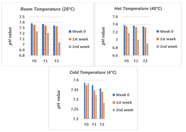
Stability tests were conducted by placing the formulas in various temperature conditions, including cold (4°C), room (28°C), and hot (40°C) temperatures, for 2 weeks, with evaluations performed every week. Observations were made on organoleptic and pH properties. The results of organoleptic observations of the three liposome preparation formulas at cold temperatures (4°C), room temperature (28°C), and hot temperatures (40°C) were conducted over 2 weeks (see Figure 4).
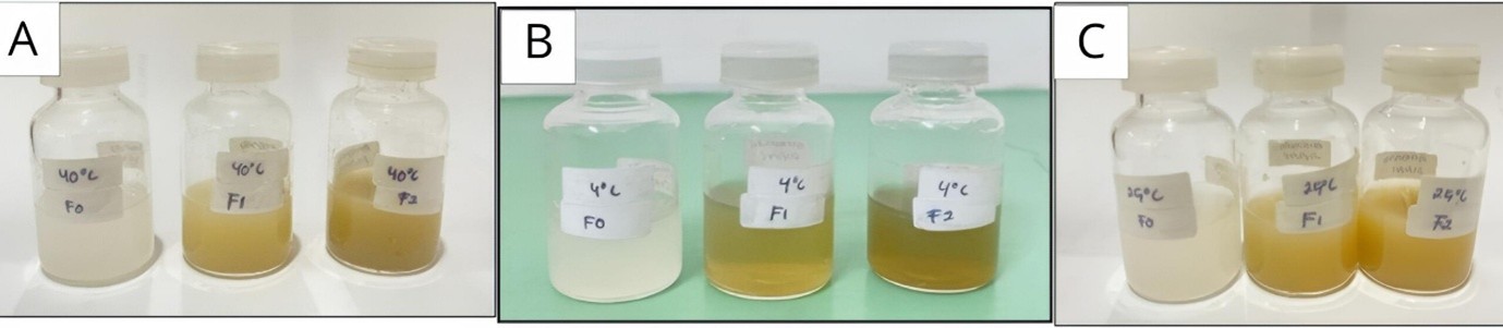
The stability test results indicated that all liposome formulations were stable only when stored at cold temperatures (4 °C). However, at room temperature (28 °C) and elevated temperatures (40 °C), signs of sedimentation and changes in pH were observed over a 14-day period. These findings suggest that the physical stability of liposomes is still limited and do not support claims of improved long-term stability. Therefore, additional tests, such as zeta potential measurements and drug leakage profiles, are necessary to quantitatively assess colloidal stability. Furthermore, real-time stability tests over a duration of 1 to 3 months will help determine actual storage performance, which is characterized by the formation of sediment (32). As explained in the research, the niosome formula containing apigenin, a flavonoid group compound, was also stable in 4°C storage for 3 months.
Based on the pH measurement results of the C. asiatica(L. ) Urban extracts liposomes after a 2-week stability test (see Figure 5). The pH of the C. asiatica(L. ) Urban extract liposomes are generally still within the standard range of oral and topical use of 4.5-8.0. However, the change in pH value at each week indicates the reaction of damage to the constituent components in the preparation. Therefore, the change in pH value will affect the effect given by the liposome (33). Such pH variations may reflect gradual chemical interactions between the lipid components and the encapsulated extract during storage. Although still within acceptable limits, continuous pH monitoring is essential to ensure formulation consistency and safety over prolonged storage periods.
DPPH method antioxidant activity test
Antioxidant activity test using the DPPH (2, 2-diphenyl-1-picrylhydrazyl) method. This method can be used to determine antioxidant activity based on the ability to counteract the DPPH radical. The maximum wavelength was obtained using a methanol and DPPH solvent mixture with a ratio of 4: 3, yielding a maximum wavelength at 515 nm with an absorbance of 0.852 and an operating time of 16 min. Operating time is used to determine the stability of the solution. To determine the antioxidant activity, it can be done by calculating the inhibitory concentration. (IC50).
The results of the antioxidant activity test of the ethanol extract of C. asiatica(L. ) Urban with F1 and F2 showed that F1 and F2 had almost the same antioxidant activity as the pure extract (see Table 7). While the IC₅₀ values of F1 and F2 were not significantly different (p > 0.05) from those of the pure extracts, suggesting that encapsulation preserved their antioxidant activity, there is currently no data connecting this retention of antioxidant activity to the controlled release profile. Furthermore, the diffusion test was conducted for only 4 h, so we cannot draw any conclusions about its biological relevance to the chronic wound healing process. The free radical scavenging ability of an extract is related to its phenol content (34).
| Sample | IC50 value | Category |
|---|---|---|
| Vitamin C | 9.16 ± 0.06 | Very strong |
| C. asiatica (L. ) Urban extract | 13.87± 0.02* | Very strong |
| Formula I | 14.10 ± 0.04* | Very strong |
| Formula II | 13.97 ± 0.06* | Very strong |
| p-value < 0, 05 vs Vit C | ||
Liposome Gel Preparation Results
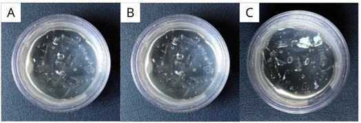
The composition of the liposomal gel formulation included triethanolamine as a neutralizing agent, propylene glycol as a humectant, and Carbopol as a gelling agent. The use of Carbopol as the gel base resulted in a formulation with superior consistency and more optimal release of active substances compared to other gelling agents (35). These characteristics indicate that Carbopol is suitable for liposomal gel formulations intended for topical application. Visually, the liposomal gel preparations containing ethanol extract exhibited a homogeneous and stable appearance, as shown in Figure 6, which includes the base gel, formulation gel 1 (FG1), and formulation gel 2 (FG2). The physical characteristics of the liposomal gel preparations, including quality parameters, are presented in Table 8 and were evaluated in accordance with applicable pharmacological guidelines.
| Parameters | Base | FG1 | FG2 |
|---|---|---|---|
| Organoleptic | Color: ClearTexture: Non-stickySmell: Typical Carbopol | Color: ClearTexture: Non-stickySmell: Typical Carbopol | Color: ClearTexture: Non-stickySmell: Typical Carbopol |
| Homogeneity | Homogeneous | Homogeneous | Homogeneous |
| Spreadability | 6.02 ± 0.072 | 6.16 ± 0.38 | 6.33 ± 0.27 |
| pH | 5.08 ± 0.04 | 5.30 ± 0.02 | 5.54 ± 0.10 |
| Viskositas | 48500.0 ± 330 | 46533.3 ± 438.444 | 33466.7 ± 1171.893 |
| flow properties | thiksotropik | thiksotropik | thiksotropik |
Based on the results of testing the characteristics of liposomal gel preparations, it can be concluded that the preparation meets the requirements of a good gel preparation. According to (36), the expected characteristics of gel preparations are a semi-solid consistency and an organoleptically transparent color, free of coarse grains or ingredients that have not mixed well, which can form lumps or be homogeneous (37).
The pH measurement was performed in triplicate for each formulation, and the average values were within the desired range of 4.5–6.5, which corresponds to the physiological pH of the skin. Maintaining the pH within this interval is critical, as overly acidic preparations may lead to irritation, while alkaline formulations can disrupt the skin barrier, causing dryness and flaking. Recent studies have emphasized that topical products should ideally match the skin’s natural acidity (approximately pH 4.5–5.5); deviations towards alkalinity increase surface pH, compromise barrier integrity, and are associated with irritation and xerosis (38). Similarly, recent emulgel formulation studies confirmed that pH values between 5.6 and 6.8 are compatible with skin physiology. They highlighted that formulations outside this range pose a greater risk of irritation and barrier dysfunction (39). Both formulations maintain a physiological pH (5.08–5.54), promoting barrier function and keratinocyte migration, essential for wound resurfacing. In conclusion, the C. asiatica liposomal gel system presents a promising approach for improving wound healing through controlled antioxidant delivery and enhanced bioavailability.
Each formula has a different diameter, as indicated by the results of the spreadability test. The results of this spreadability can be influenced and are inversely proportional to the viscosity of a preparation (40). To ensure the observation results obtained are accurate, namely that the greater the concentration of liposomes in the formula, the greater the spreadability increases. Therefore, when the viscosity increases, the ability to spread a preparation on the skin surface decreases (41). According to (SNI 16-4380-1996), the requirement for viscosity value in gel preparations is 3000-50000 cPs (42).
The determination of flow properties in each formula reveals thixotropic flow. This indicates that the preparation has a lower viscosity value at each shear velocity value of the decreasing curve than in the increasing curve (43). Thixotropic flow properties are expected in gel preparations, where the preparation exhibits high consistency in the container but can be easily removed with little force and spreads smoothly when applied to the skin (44). Thus, when applied to the skin, the gel spreads easily (43). This rheological behavior contributes to improved user comfort and uniform application during topical use. Additionally, thixotropy may help maintain formulation stability by reducing structural breakdown during storage. The observed thixotropic flow indicates that the gel exhibits reduced viscosity under shear, facilitating easy spreading during application while maintaining consistency at rest. This rheological behavior supports practical handling and uniform topical application without implying additional functional effects.
In-Vitro Penetration Test
The purpose of the penetration test was to determine the ratio of quercetin penetration from the liposome gel with varying liposome concentrations through the skin during a specific time interval. The test was conducted for 4 h, and sampling was done at 6 points, namely at the 30th, 60th, 90th, 120th, 180th, and 240th min. The in vitro percutaneous penetration test has two main parameters: the cumulative amount of penetrated active substance or percentage of penetrated dose, and the penetration rate or flux (45).
The standard curve of quercetin in phosphate buffer at pH 7.4 was obtained at a wavelength of 374.5 nm: y = 0.0196x - 0.0557, with R² = 0.9982. Figure 7 presents the cumulative permeation of quercetin through rat skin from liposomal gels over 4 h. FG2 achieved 314.40 μg/cm², exceeding FG1 (280.86 μg/cm²) by ≈11.9%. The steady-state permeation rates confirmed this advantage: 79.741 ± 0.421 μg/cm²·h for FG2 versus 67.234 ± 1.439 μg/cm²·h for FG1. The superior performance of FG2 is attributable to its smaller particle size (101.3 nm) and lower PDI (0.306) relative to FG1 (119.8 nm; PDI = 0.410), as smaller and more monodisperse liposomes exhibit improved diffusion through the gel matrix and enhanced skin partitioning (46). Consistent with transdermal delivery principles, liposomes < 300 nm penetrate beyond the stratum corneum more effectively, whereas larger vesicles show limited depth of delivery (2). These data indicate that optimizing vesicle size and PDI is a practical lever to increase quercetin flux and total delivery in early time windows (47). The results of the drug release kinetics model determination for the liposomal gel formulations are presented in Table 9.
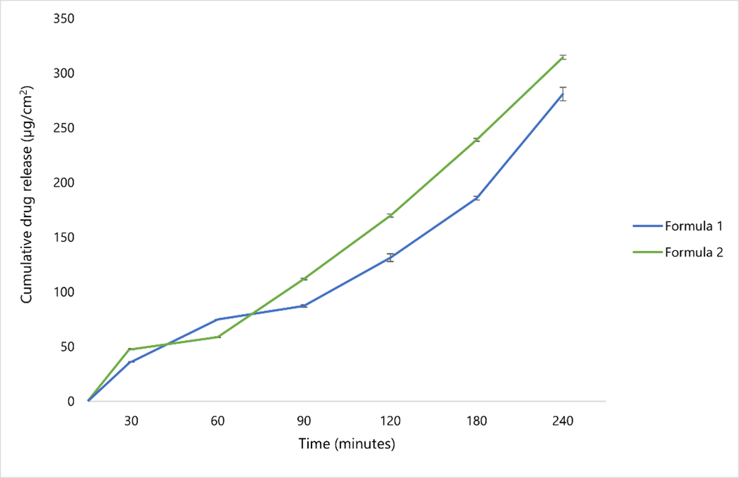
Table 9. Results of drug release kinetics model determination.
| Formula | Orde Nol | Orde 1 | Higuchi | Kosmeyer-peppas |
|---|---|---|---|---|
| F1 | 0.9881 | 0.6211 | 0.8745 | 0.8799 |
| F2 | 0.9914 | 0.6229 | 0.8859 | 0.8799 |
The analysis of release kinetics revealed that the data fit best to the zero-order model, with R² values ranging from 0.9881 to 0.9914. However, relying solely on the R² value is insufficient to confirm a pure zero-order release mechanism. When compared to the Higuchi model (R² = 0.8745-0.8859) and the Korsmeyer-Peppas model (R² = 0.8799), there is evidence of matrix diffusion contributing to the release process. Therefore, the release mechanism is more accurately described as a mixed (anomalous) release, dominated by reservoir components. To fully validate these mechanistic conclusions, further residual studies and mathematical modeling are necessary. Similar findings have been reported for quercetin microemulsion gels and other vesicular-hydrogel systems, where optimized matrices flatten release curves and prolong delivery (48,49). Overall, the release profile indicates a controlled and gradual release pattern rather than a single ideal kinetic model. The higher R² values in the zero-order model suggest a near-constant release rate, while the contribution of diffusion-based mechanisms reflects the role of the formulation matrix. This combination is commonly observed in vesicular or hydrogel-based systems with structured matrices. Consequently, the release behavior can be described as consistent over time without implying enhanced efficacy. In addition, the in vitro penetration and release data provide comparative insight into the influence of liposomal characteristics on transdermal transport behavior under controlled conditions. The observed differences between formulations reflect formulation-driven optimization rather than pharmacodynamic enhancement. These findings support the use of in vitro penetration and release assessments as preliminary screening tools for evaluating compounds before further biological or in vivo evaluation.
Conclusion
This study developed liposomal gels incorporating ethanol extract of C. asiatica, showing favorable physicochemical properties, preserved antioxidant activity, and improved skin penetration. The optimized formulation, FG2 (0.5%), had a particle size of approximately 101 nm and a low polydispersity index (PDI) of 0.306, indicating homogeneous vesicle distribution.
Both FG1 and FG2 achieved strong antioxidant activity (IC₅₀ ≈ 13.9–14.1 µg/mL) and enhanced skin permeation compared to FG1 over 240 min. Kinetic analysis suggested a zero-order release model (R² > 0.99), though further modeling is needed to confirm the release profile. Stability was observed only at 4 °C, suggesting the need for further optimization of lipid composition. These findings highlight the potential of liposomal gels as carriers for topical antioxidant delivery, relevant for wound healing. Future research should focus on long-term stability, zeta-potential, encapsulation efficiency, and in vivo wound healing performance for improved therapeutic applications.
Abbreviations
CA = C. asiatica; FG = Formula Gel; PS = Particle Size; PDI = Polydispersity Index; ZP = Zeta Potential; EE% = Entrapment Efficiency Percentage; DPPH = 2, 2-Diphenyl-1-picrylhydrazyl; IC50 = Inhibitory Concentration 50%; SD = Standard Deviation; r² = Coefficient of Determination; FHI = Farmakope Herbal Indonesia (Indonesian Herbal Pharmacopoeia); UV-Vis = Ultraviolet–Visible Spectrophotometry.
Declarations
Acknowledgment
The authors would like to express their sincere gratitude to the Ministry of Education, Culture, Research, and Technology of the Republic of Indonesia (Kemendikbudristek) for funding this study through the Beginner Lecturer Research Grant (PDP), managed under the Directorate General of Higher Education, Research, and Technology and coordinated by the Directorate of Research and Community Service (DPPM).This work was funded under Contract Number 125/C3/DT.05.00/PL/2025, 7964/LL4/PG/2025, and 677/II.3.UMMADA/F/2025. The authors also gratefully acknowledge University of Muhammadiyah Ahmad Dahlan Cirebon, the Institute for Research and Community Service (LPPM), the Faculty of Pharmacy, and the Pharmacy Study Program for their continuous support, facilities, and encouragement that greatly contributed to the successful completion of this research and publication.
Conflict of Interest
The authors declare no conflicting interest.
Data Availability
All data generated or analyzed during this study are included in this published article
Ethics Statement
All animal experiments were approved by the [health research ethics committee, University Muhammadiyah Ahmad Dahlan cirebon] (Approval No. 002/VII/2025/0004/KEPK/STFMC) and conducted in accordance with relevant guidelines and regulations.
Funding Information
The authors would like to express their sincere gratitude to the Ministry of Education, Culture, Research, and Technology of the Republic of Indonesia (Kemendikbudristek) for funding this study through the Beginner Lecturer Research Grant (PDP), managed under the Directorate General of Higher Education, Research, and Technology and coordinated by the Directorate of Research and Community Service (DPPM).This work was funded under Contract Number 125/C3/DT.05.00/PL/2025, 7964/LL4/PG/2025, and 677/II.3.UMMADA/F/2025.
References
- Sen CK. Human wounds and its burden: an updated compendium of estimates. Adv Wound Care (New Rochelle). 2019;8(2):39–48.
- Jain S, Patel N, Shah MK, Khatri P, Vora N. Recent advances in lipid-based vesicles and particulate carriers for topical and transdermal application. J Pharm Sci. 2017;106(2):423–45.
- Witkowska K, Paczkowska-Walendowska M, Garbiec E, Cielecka-Piontek J. Topical application of Centella asiatica in wound healing: recent insights into mechanisms and clinical efficacy. Pharmaceutics. 2024;16(10):1252.
- Mirza MA, Mahmood S, Hilles AR, Ali A, Khan MZ, Zaidi SAA, et al. Quercetin as a therapeutic product: evaluation of its pharmacological action and clinical applications—a review. Pharmaceuticals. 2023;16(11):1631.
- Rosalina AI, Sagita E, Iskandarsyah. Penghantaran obat melalui kulit: teknologi vesikel liposom dan analognya. J Kedokt Meditek. 2023;29(1):109–20.
- Rivera AD, Pieropan F, Williams G, Calzolari F, Butt AM, Azim K. Drug connectivity mapping and functional analysis reveal therapeutic small molecules that differentially modulate myelination. Biomed Pharmacother. 2022;145:112436.
- De Luca M, Tuberoso CIG, Pons R, García MT, Morán MDC, Martelli G, et al. Ceratonia siliqua L. pod extract: from phytochemical characterization to liposomal formulation and evaluation of behaviour in cells. Antioxidants. 2023;12(6):1209.
- Mirshekari M, Bagheri Ghomi A, Hamishehkar H, Farahpour MR. In vivo evaluation of wound healing activity of nanoliposomes loaded Withania somnifera extract. Adv Pharm Bull. 2024;14(4):846.
- Elmaidomy AH, Mohamad SA, Abdelnaser M, Yahia R, Mokhtar FA, Alsenani F, et al. Vitis vinifera leaf extract liposomal Carbopol gel preparation: potential wound healing and antibacterial benefits. Food Funct. 2023;14(15):7156–75.
- Thorat YS, Kote NS, Patil VV, Hosmani AH. Formulation and evaluation of liposomal gel containing extract of piperine. Int J Curr Pharm Res. 2020;12(4):31–6.
- Chaerunisaa AY, Dewi MK, Sriwidodo, Joni IM, Dwiyana RF. Development of cathelicidin in liposome carrier using thin-layer hydration method. Int J Appl Pharm. 2022;14(4):178–85.
- Lombardo D, Kiselev MA. Methods of liposome preparation: formation and control factors of versatile nanocarriers for biomedical and nanomedicine application. Pharmaceutics. 2022;14(3):543.
- Shahidi F, Zhong Y. Measurement of antioxidant activity. J Funct Foods. 2015;18:757–81.
- Baliyan S, Mukherjee R, Priyadarshini A, Vibhuti A, Gupta A, Pandey RP, et al. Determination of antioxidants by DPPH radical scavenging activity and quantitative phytochemical analysis of Ficus religiosa. Molecules. 2022;27(4):1326.
- Ng SF, Rouse JJ, Sanderson FD, Meidan V, Eccleston GM. Validation of a static Franz diffusion cell system for in vitro permeation studies. AAPS PharmSciTech. 2010;11(3):1432–41.
- Azmin M, Siti Nuurul Huda, Mat Nor MS. Chemical fingerprint of Centella asiatica bioactive compounds in ethanolic and aqueous extracts. Adv Biomark Sci Technol. 2020;2:35–44.
- Departemen Kesehatan Republik Indonesia. Farmakope Indonesia. 2nd ed. Jakarta; 2017.
- Fitriyanti F, Qalbiyah S, Sayakti P. Identifikasi kulit batang kalangkala (Litsea angulata BI) secara makroskopik, mikroskopik, dan skrining fitokimia. Parapemikir J Ilm Farm. 2020;9(2):1–9.
- Kandasamy A, Aruchamy K, Rangasamy P, Varadhaiyan D, Gowri C, Oh TH, et al. Phytochemical analysis and antioxidant activity of Centella asiatica extracts. Plants. 2023;12(20):3547.
- Rashid MdHO, Akter MstM, Uddin J, Islam S, Rahman M, Jahan K, et al. Antioxidant, cytotoxic, antibacterial and thrombolytic activities of Centella asiatica L. Clin Phytosci. 2023;9(1):1.
- Mewar D, As’ad MuhF. Standarisasi parameter spesifik dan non-spesifik ekstrak etanol daun gatal (Laportea decumana (Roxb.) Wedd). J Penelit Kesehat SUARA FORIKES. 2023;14(2).
- Mohapatra P, Ray A, Jena S, Nayak S, Mohanty S. Influence of extraction methods and solvent system on the chemical composition and antioxidant activity of Centella asiatica L. leaves. Biocatal Agric Biotechnol. 2021;33:101971.
- Balekundri A, Mannur V. Quality control of the traditional herbs and herbal products: a review. Futur J Pharm Sci. 2020;6(1):67.
- Shraim AM, Ahmed TA, Rahman MM, Hijji YM. Determination of total flavonoid content by aluminum chloride assay: a critical evaluation. LWT. 2021;150:111932.
- Chandra S, Khan S, Avula B, Lata H, Yang MH, ElSohly MA, et al. Assessment of total phenolic and flavonoid content, antioxidant properties, and yield of aeroponically and conventionally grown crops. Evid Based Complement Alternat Med. 2014;2014:1–9.
- Widyasari EM, Sriyani ME, Daruwati I, Halimah I, Nuraeni W. Karakteristik fisikokimia senyawa bertanda 99mTc-kuersetin. J Sains Teknol Nukl Indones. 2019;20(1):9.
- Solikah WY, Fatmawati A, Gunawan A, Defri AY. Uji kualitatif dan penetapan kadar flavonoid total ekstrak etanol herba pegagan (Centella asiatica). J Pharm Sci. 2023;6(2):673–80.
- Khairunnisa S, Hakim AR, Audina M. Perbandingan kadar flavonoid total ekstrak daun pegagan (Centella asiatica (L.) Urban). J Pharm Care Sci. 2022;3(1):121–31.
- Desai J, Mallya R. Development of green coffee beans extract loaded anti-aging liposomal gel. Indian J Pharm Educ Res. 2021;55(4):979–88.
- Apriani DK, Dwiya RF, Chaerunisaa AY, Rostinawati T. Pengembangan peptida antimikroba sintesis (KR-12) dalam sistem vesikular liposom. Maj Farmasetika. 2024;9(2):179.
- Indalifiany A, Suryani S, Aspadiah V, Mahmudah R, Annisa R. Formulasi gel dari niosom elastik ekstrak Etlingera alba. J Sains Kesehat. 2023;5(5):675–84.
- Mujtaba MdA. Development of apigenin-loaded niosomes using ecological probe sonication technique. Curr Pharm Biotechnol. 2022;23(6):882–93.
- Putra MM, Dewantar IGNA, Swastini DA. Pengaruh lama penyimpanan terhadap nilai pH sediaan cold cream kombinasi ekstrak kulit buah manggis (Garcinia mangostana L.), herba pegagan (Centella asiatica) dan daun gaharu (Gyrinops versteegii (Gilg) Domke). J Farm Udayana. 2014;3(1):279745.
- Widyani M, Ulfa M, Wirasisya DG. Efek penghambatan radikal bebas herba pegagan (Centella asiatica). J Pijar MIPA. 2019;14(1):100–6.
- Andika BT, Rahmawati D, Kuncoro H. Uji aktivitas antioksidan dan formulasi gel ekstrak Citrus aurantifolia. Proc Mulawarman Pharm Conf. 2021;14:25–30.
- Putri WE, Anindhita MA. Optimization of cardamom fruit ethanol extract gel. J Ilm Farm. 2022:107–20.
- Safitri CINH, et al. Formulasi dan uji mutu fisik sediaan gel ekstrak bekatul (Oryza sativa L.). In: Prosiding Seminar Nasional Pendidikan Biologi dan Saintek (SNPBS). 2020:228–235.
- Lukić M, Pantelić I, Savić SD. Towards optimal pH of the skin and topical formulations. Cosmetics. 2021;8(3):69.
- Mahmood A, Erum A, Tulain UR, Malik NS, Saleem A, Alqahtani MS, et al. Exploring the gelling properties of Plantago ovata-based arabinoxylan. PLoS One. 2023;18(8):e0290223.
- Indah NHS. Formulasi sediaan serum kosmetik kombinasi ekstrak kelopak bunga rosela merah (Hibiscus sabdariffa L.) dengan minyak biji bunga matahari (Helianthus annuus L.) serta uji aktivitas antioksidan dengan metode DPPH. Thesis. Universitas Sumatera Utara; 2021.
- Pratiwi L, Wahdaningsih S. Formulasi dan aktivitas antioksidan masker wajah gel Carica papaya. J Farm Medica. 2018;1(2).
- Pertiwi RD, Kristanto J, Praptiwi GA. Uji aktivitas antibakteri gel ekstrak Abrus precatorius. J Ilm Manuntung. 2017;2(2):239–47.
- Chandra D, Fitria. Formulasi sediaan gel, krim, dan gel-krim ekstrak Coffea arabica. J Ilm Farm Imelda. 2019;2(2):45–50.
- Pricillya LM, Falestin SLK, Julisna S. Formulasi gel ekstrak Zingiber officinale var. rubrum. J Ris Kefarm Indones. 2019;1(2).
- Yani TN, Anwar E, Saputri FC. Formulasi emulgel ekstrak Anredera cordifolia. J Kefarm Indones. 2016;6(2):88–97.
- Danaei M, Dehghankhold M, Ataei S, Hasanzadeh Davarani F, Javanmard R, Dokhani A, et al. Impact of particle size and polydispersity index on lipidic nanocarriers. Pharmaceutics. 2018;10(2):57.
- Abd E, Gomes J, Sales CC, Yousef S, Forouz F, Telaprolu KC, et al. Deformable liposomes as enhancer of caffeine penetration. Int J Cosmet Sci. 2021;43(1):1–10.
- Laracuente ML, Yu MH, McHugh KJ. Zero-order drug delivery: state of the art and future prospects. J Control Release. 2020;327:834–56.
- Kajbafvala A, Salabat A, Salimi A. Formulation and evaluation of quercetin-loaded microemulsion for topical application. Pharm Dev Technol. 2018;23(8):741–50.