RESEARCH ARTICLE
Phytochemical Screening and Antioxidant Activity of Mimosa tenuiflora (Willd.) Poir Root Extracts
Academic Editor: Emad Mohamed Abdallah
Sciences of Phytochemistry|Vol. 5, Issue 1, pp. 52-63 (2026)
Views
Downloads
Shares
Received
Nov 28, 2025Revised
Feb 9, 2026Accepted
Mar 5, 2026Published
Apr 6, 2026
Abstract
This study evaluated the phytochemical profile and antioxidant activity of extracts and partitions derived from the root of Mimosa tenuiflora (Willd. ) Poir. , also known as tepezcohuite. Three extracts were prepared through hydroalcoholic maceration, total alkaloid extraction, and Soxhlet extraction. Additionally, four partitions were obtained from the ethanolic extract using the modified Kupchan technique: n-hexane, dichloromethane, n-butanol, and water. Qualitative phytochemical screening showed the presence of alkaloids, tannins, flavonoids, saponins, steroids, triterpenoids, reducing sugars, coumarins, and cardiac glycosides in various several fractions, while no anthraquinone glycosides were detected. Antioxidant activity was assessed using DPPH and ABTS assays. In the DPPH assay, the most active fractions were the butanolic (EC50 = 2.20 ± 0.45 µg/mL) and the ethanolic (EC50 = 2.25 ± 0.01 µg/mL). While, in the ABTS assay, the ethanolic extract and butanol partition were the most active fractions with EC50 values of 4.87 ± 1.11 µg/mL and 5.43 ± 0.21 µg/mL respectively. The less polar fractions (n-hexane, Soxhlet) exhibited lower activity. This study expands the phytochemical knowledge of M. tenuiflora, focusing on its roots, an organ less extensively characterized than the bark regarding its comprehensive phytochemical and antioxidant profile. The results show that this organ is a promising source of bioactive metabolites with antioxidant capacity. This finding justifies further investigation into the pharmacological and therapeutic applications of these compounds, which is particularly relevant given that while previous research has systematically favored the bark, and existing root studies have focused primarily on specific alkaloids, comprehensive profiling remains limited.
Keywords:
Introduction
Mimosa tenuiflora (Willd.) Poir., commonly known in Mexico as tepezcohuite, is a native American plant belonging to the Fabaceae family. It’s distributed in areas of deciduous tropical rainforest, ranging from southeastern Mexico (Oaxaca and Chiapas) to Honduras, El Salvador, Panama, Colombia, Venezuela, and as far north as northern Brazil. It is a thorny evergreen tree that can reach heights of up to 8 meters. It has alternate compound leaves with 5-10 pairs of pinnae and 10-30 pairs of linear leaflets, white flowers grouped in dense spikes, 3-6.5 cm long, and a lanceolate, unarmed fruit, compressed between the seeds, 2-4.5 cm long and 5-7 mm wide, with 2-6 segments. Young branches, leaves, and immature fruits have glandular hairs and/or glands (1).
From an ethnobotanical perspective in Mexico, the use of this plant has had significant relevance since the 1980s. The tree bark has been used for burning and dermatological conditions, specifically mild to severe burns, and reduce inflammation (2–4). Regarding other studies on biological activity, it has been shown that extracts obtained from the tree bark exhibit antiseptic (5) and antimicrobial properties (6), antifungal activity (7), inhibitory properties in the aflatoxins production (8), and antioxidant activity. The evaluation of antioxidant activity has focused mainly on bark extracts. Magalhães et al., 2018, reported that the ethanolic extract of this fraction showed the highest antioxidant activity (EC₅₀ = 132.99 μg/mL in DPPH and 189.14 μg/mL in ABTS) (9). While López et al., 2022 observed considerably lower activity for an ethanolic extract (IC₅₀ = 464.06 ± 8.58 µg/mL in DPPH), especially compared to ascorbic acid (IC₅₀ = 2.23 ± 0.42 µg/mL) (10). In contrast, research on the antioxidant activity of root extracts within the Mimosa genus remains scarce, with previous studies focusing predominantly on species such as Mimosa hamata Willd. and Mimosa pudica L. (11, 12). While recent metabolomic profiles of M. tenuiflora root bark have been reported by de Sousa et al., 2024 (13), comprehensive studies linking solvent polarity partitions to specific antioxidant endpoints using standardized benchmarks such as trolox and quercetin are still limited. This restricted approach highlights a critical gap in the phytochemical and antioxidant characterization of M. tenuiflora roots, specifically regarding the distribution of activity across polarity fractions. Hitherto, studies about the root have been mainly limited to the extraction of a primary alkaloid with psychotropic activity, this being N, N-dimethyltryptamine (14, 15), just like an anxiolytic potential based on the presence of this same alkaloid (16) and as an alternative for the phytopathogen control (17). While this raises important regulatory and toxicological considerations, the presence of co-occurring non-alkaloid metabolites warrants investigation for potential therapeutic applications, provided safety profiles are established in future studies.
Therefore, this study aimed to obtain extracts and partitions from the root of M. tenuiflora using hydroalcoholic, total alkaloid, and Soxhlet methods, followed by simplification of the ethanolic extract using the modified Kupchan method to isolate partitions of varying polarity. Additionally, phytochemical screening was performed for the preliminary identification of secondary metabolites. Finally, the median effective concentration (EC₅₀) was determined via DPPH and ABTS assays, establishing comparability with trolox and quercetin reference standards as the objective criterion for relevant antioxidant activity.
Experimental Section
Materials
Reagents
The reagents and solvents employed during this study were of analytical grade, and solutions were prepared using distilled water. There are: α-Naphthol (99% purity), ammonium hydroxide (28–30% purity), cholesterol (99% purity), chloroform (99% purity), ferric chloride (97% purity), hydrochloric acid (36.5% purity), mercuric chloride (95.5% purity), magnesium metal (Mg), picric acid, potassium iodide (99% purity), saponin (99% purity), sodium carbonate (99% purity), sodium hydroxide (97% purity) (Fermont, Mex). Acetic anhydride (97% purity), copper sulphate (99.9% purity), dichloromethane (99% purity), glacial acetic acid (99.7% purity), sodium citrate (99.9% purity), and sulfuric acid (95–98% purity) (J. T. Baker, USA). 2, 2′-Azino-bis (3-ethylbenzothiazoline-6-sulfonic acid) diammonium salt (ABTS) (98% purity), 2, 2-diphenyl-1-picrylhydrazyl (DPPH•) (97% purity), atropine sulfate salt monohydrate (97% purity), carminic acid (90% purity), digoxin (95% purity), fructose (99% purity), quercetin (95% purity), sodium nitroprusside (99% purity), trolox (97% purity), vanillin (99% purity) (Sigma-Aldrich, USA). Ethanol (99.5% purity), methanol (99.8% purity) (Jalmek, Mex). 4-(Dimethylamino)benzaldehyde (98% purity), acetone (99% purity), iodine (I₂) (99% purity), n-butanol (99% purity), n-hexanes (98% purity), petroleum ether (95% purity), potassium persulfate (99% purity) (Golden Bell, Mex). Bismuth (III) nitrate pentahydrate (Bi (NO₃)₃·5H₂O) (98.9% purity), gallic acid (98.7% purity), gelatin (Fagalab, Mex). Sodium chloride (99.9% purity) (CDH, IN), and coumarin (MC/B, USA).
Plant Material
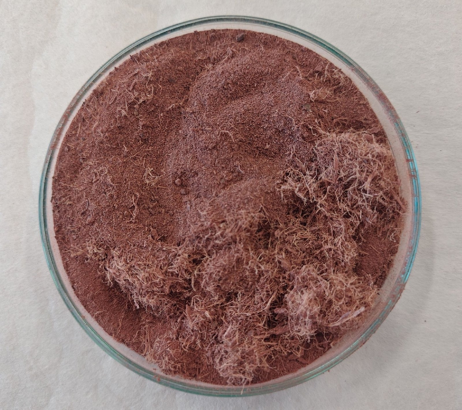
The root of M. tenuiflora was purchased online through the Mercado Libre Mexico commercial platform (mercadolibre. com. mx) under the brand name Benlaifh, a supplier of natural products. It was collected in the community of Cintalapa de Figueroa, Chiapas, Mexico. Due to the commercial nature of the acquisition, no voucher specimen was deposited in an herbarium. The root appears as an intense reddish-brown powder with a fibrous texture, a characteristic smell, slightly resinous aroma, and a slightly bitter, astringent taste (Figure 1). Once obtained, the material was stored at -70 °C until use. It is noteworthy that the raw root material of M. tenuiflora is not classified as a controlled or illicit substance under current Mexican federal legislation.
Instrumentation
Microplate reader spectrometer Epoch (BioTek Instruments, Inc., USA), Büchi Rotavapor® R-100 (Büchi, CH) and Ultrasonic cleaner Cole-Parmer® model 08895-05 (Cole-Parmer, USA).
Preparation of extracts
Hydroalcoholic Maceration
300 g of botanical material was subjected to cold alcoholic extraction with 700 mL of ethanol and distilled water in a 7:3 ratio. This mixture is shaken occasionally for 5 days at a constant temperature of 20 °C and kept away from direct sunlight. Later, the extract was filtered by gravity using a funnel and filter paper. Then, the extract was evaporated under reduced pressure with a Rotavapor® apparatus equipped with a heating bath at 35 °C until dryness. The solvent removed by vacuum was used to perform the maceration again. This process was repeated twice.
Total Alkaloids Extraction
The total alkaloid extraction was carried out using the methodology proposed by Rossi et al., 2019 with some modifications (14). Briefly, 300 mL of distilled water and 40 g of NaOH were added to a beaker with a magnetic stirrer. Once dissolved, 80 g of NaCl was incorporated and allowed to dissolve. The solution was then heated to 70 °C. Separately, in an Erlenmeyer flask with a magnetic stirrer, 100 g of M. tenuiflora root was rehydrated in 200 mL of distilled water. When hydrated and under constant stirring, the NaOH/NaCl solution was added, resulting in a dark-brown suspension. It was stirred and maintained at 50-55 °C for 10 minutes. After adding 200 mL of n-hexanes, the mixture was vigorously stirred for another 10 minutes. The stirring was then stopped, and the phases were allowed to separate. The organic phase was separated and evaporated under reduced pressure using a Rotavapor® apparatus until all solvents were removed. The n-hexanes extraction was repeated twice on the basic mixture.
Soxhlet Extraction
The Soxhlet system and a borosilicate thimble were pre-washed with a 1:1 mixture of n-hexanes and acetone before use. Afterward, the material was left in a heating oven for 4 hours until it was completely dry. 50 g of finely ground plant material was weighed and placed in the borosilicate thimble inside the Soxhlet extractor. 350 mL of petroleum ether served as the extraction solvent. The system was coupled to a 500 mL round-bottom flask and heated under reflux at a temperature between 65 and 70 °C for 5 hours using a glycerol double boiler. Then, the extract was concentrated under reduced pressure using a Rotavapor®.
Modified Kupchan’s Solvent-Partitioning Method
The dried ethanolic extract was subjected to a solvent partitioning process, as described by Kupchan (1980) with modifications, which serves to separate the different components present in the plant extract based on their solubility differences (18). 100 g of the dried extract was rehydrated with 100 mL of ethanol, then mixed with 300 mL of distilled water and stirred until completely dissolved. The mixture was transferred to a separatory funnel, where 300 mL of n-hexanes were added. After constant stirring (3 min, with venting intervals) to prevent emulsion formation, the mixture was left to stand until phase separation occurred (about 5 minutes), and the phases were collected separately. The residual water/ethanol phase was returned to the funnel, and the process was repeated with 300 mL of dichloromethane, stirring gently (for 3 min with venting intervals, avoiding emulsion formation, and waiting approximately 10 minutes for phases to separate). Each partition was collected separately. The remaining aqueous/ethanol phase was partitioned with 300 mL of n-butanol using the same procedure (3 min with venting intervals, avoiding emulsion formation, and roughly 35 minutes for phase separation). Each step used a total of 900 mL of solvent, divided into 300 mL portions.
Storage Conditions for Extracts and Partitions
The extracts and their partitions were evaporated to dryness under controlled conditions and weighed periodically until a constant weight was reached. They were then stored at -20 °C in airtight containers under N2 atmosphere protected from light to preserve their chemical integrity until further analysis.
Qualitative Analysis of Phytochemical Screening
To identify the secondary metabolites, present in the extract and partitions obtained, an exhaustive phytochemical screening was performed using specific colorimetric assays. Each assay was performed in triplicate (n = 3) using at least two independent analytical methods per metabolite class (except anthraquinone glycosides), with aliquots prepared independently to ensure reproducibility. As experimental controls, a negative blank (solvent + corresponding reagent) and a positive control (specific reference standard + reagent) were included, which allowed the stability of the colorimetric reagents and the validity of the observed responses to be confirmed. Qualitative observations were interpreted by systematic comparison with the positive control reactions and reactivity patterns described in the specialized literature (19–24). For more details on quantities and volumes used, please consult the supplementary material (Annex 1).
Determination of Alkaloids
An aliquot of dry weight of the samples to be evaluated was diluted in 5% HCl, and sonicated until completely dissolved. Subsequently, the aliquots were taken and placed in test tubes (one for each test). Then, a few drops of each corresponding reagent for the test were added, shaken lightly, and allowed to stand for 1 to 2 minutes. A positive result was considered when: Mayer: cream-colored precipitate, Wagner: red-brown, brown, or orange precipitate, Dragendorff: reddish-brown or brown precipitate and Hager: yellow precipitate or yellow suspension accompanied by a reddish precipitate.
Determination of Tannins
An aliquot of the test solution was placed in a test tube, followed by the addition of a few drops of each reagent corresponding to each test. For the FeCl3 test, a positive result was indicated by the formation of a wine-red, intense green, blue, dark blue, or black color. For the gelatin test, the appearance of a white precipitate was considered positive.
Determination of Flavonoids
Ammonia vapor reaction: A strip of filter paper was soaked in the tested solution, allowed to dry at room temperature, and then exposed to ammonia vapor. The appearance of yellow color was considered a positive sign. Shinoda test: An aliquot of the test solution was taken, and two drops of concentrated HCl were added to the walls of the tube. The reaction was then allowed to stand for five minutes, whereupon it was observed. Next, some metallic Mg filings were added to the same tube and allowed to stand for five minutes, followed by observation. The reaction is considered positive when: addition of HCl results in a red color, indicating aurones or chalcones. With the addition of Mg: orange color indicates flavones; red indicates flavonols; magenta indicates flavanones.
Determination of Steroids and Triterpenoids
Salkowski test: An aliquot of the test solution was taken, mixed with a few drops of chloroform, and two drops of concentrated H2SO4 were carefully added until a double phase was formed. The chloroform layer turned bluish red, then cherry red, and finally purple, indicating the sterols presence. If the chloroform layer changes to a reddish-brown color, it indicates the presence of a triterpenoid nucleus. Liebermann-Burchard test: An aliquot of the test solution, a few drops of acetic anhydride and two drops of chloroform were added, mixed gently, and then two drops of concentrated H2SO4 were carefully added through the walls of the tube. If the solution turns red, then blue, then blue-green, or yellow to dark green; it indicates the presence of the steroid nucleus. If the solution turns pink, red, or violet, it indicates the presence of the triterpenoid nucleus.
Determination of Saponins
Froth test: a fraction of dry analyte was added to a test tube, followed by 1 mL of distilled water. The mixture was shaken vigorously for a few minutes and then left to rest for at least 10 minutes. If a stable froth appears after 10 minutes, it indicates the presence of saponins. Rosenthaler test: a fraction of the analyte was added to a test tube, followed by three drops of the Rosenthaler's reagent, and the mixture was homogenized. Then, two drops of concentrated H2SO4 were added slowly along the walls of the tube. The formation of violet, red, and red-purple color in the middle layer was indicative of the saponins presence.
Determination of Reducing Sugars
Benedict test: An aliquot of the solution to be tested was added a few drops of Benedict's reagent. Then the tube was placed in a boiling water bath and waited for five to ten minutes. The results were interpreted according to the color change and the formation of precipitation. The persistence of the blue color indicated the absence of reducing sugars (negative result). Conversely, the appearance of a green, yellow, orange, or brick-red precipitate was interpreted as a positive result for the presence of reducing sugars. Molisch reaction: An aliquot of the solution to be tested was taken, then, a few drops of Molisch's reagent were added, and the mixture was mixed. Then, two drops of concentrated H2SO4 were slowly added without mixing to form a layer. The appearance of a purple or violet ring between the acid and the test layers indicates a positive reaction.
Determination of Anthraquinone Glycosides
An aliquot of the solution to be tested, two drops of 5% HCl were added, followed by two drops of chloroform, and finally, two drops of 10% NH₄OH. The appearance of a pink to violet ring in the ammonia phase indicates a positive result.
Determination of Cardiac Glycosides
Baljet test: An aliquot of the solution to be evaluated was taken, and two drops of the freshly prepared Baljet's reagent were added. The appearance of orange or red color indicates a positive result. Keller-Kiliani’s test: A portion of the dry analyte or an aliquot of test solution was mixed with two drops of Keller-Kiliani's reagent. To this solution, two drops of concentrated H2SO4 were added. The appearance of a blue-green, reddish-brown, brown, violet, or red ring within a few minutes indicates a positive result. Legal test: An aliquot of the test solution was dissolved in a few drops of pyridine, and two drops of 5% sodium nitroprusside solution. Finally, two drops of 20% NaOH were added through the walls of the tube. The development of pink to blood red or dark red color formation indicates positive result
Determination of Coumarins
Ehrlich test: An aliquot of the solution to be tested was taken, and few drops of fresh Ehrlich's reagent were added. The test is considered positive if a light yellow or orange color appears. NaOH test: An aliquot of the solution to be tested and two drops of 10% NaOH reagent were added. The test is considered positive if the solution turns yellow.
In vitro Antioxidant Activity Assay
The antioxidant activity of different extracts and partitions was evaluated using DPPH and ABTS radical decolorization assays. The first assay was performed following the method described by Salazar-Aranda et al., 2015 (25), with minor modifications. The second was conducted using an adapted protocol based on those described by Re et al., 1999 and Kuskoski et al., 2004 (26, 27). In both assays, quercetin and trolox (purity > 95%) served as positive controls. Each assay was performed in triplicate (n = 3). All samples were dissolved in methanol for testing. The relationship between concentration and scavenging percentage was plotted, and antioxidant activity was expressed as the median effective concentration (EC50). All experiments were conducted in triplicate using an Epoch spectrophotometer. For DPPH•, absorbance was measured at 517 nm, and for ABTS+, at 734 nm. Results are presented as mean ± standard deviation.
2, 2-Diphenyl-1-Picrylhydrazyl (DPPH) Radical-Scavenging Assay
A DPPH• solution [50 µM] was prepared and protected from light. In a 96-well plate, 200 µL of the sample to be tested was added to row A. Then, a serial dilution was made (rows B-H) by mixing 100 µL of methanol with 100 µL of the sample from row A. Finally, 100 µL of DPPH solution was added to all wells, starting from row H up to row A. The plate was then shielded from direct light and left to react for 15 minutes. The final absorbance (Af) was measured after this period. The 100% radical absorbance control (Ai) consisted of 100 µL of methanol mixed with 100 µL of DPPH• solution. The DPPH• reduction percentage was calculated using the following Eq. 1.
Where Ai represents the initial absorbance (DPPH• + methanol) and Af represents the final absorbance of the sample (DPPH• + sample).
2, 2'-Azino-bis (3-ethylbenzothiazoline-6-sulfonic acid) (ABTS) Radical-Scavenging Assay
In a 96-well plate, 24 µL of methanol was added to rows B through H, while 48 µL of the test solution was placed in row A. The serial dilution was performed by transferring 24 µL from row A to row B, and so on. The working solution was prepared by mixing 400 µL of ABTS⁺ with 16 mL of methanol. Then, 216 µL of this solution was added to each well, starting from row H and moving sequentially to row A. The plate was then shielded from direct light and allowed to react for 7 minutes. This value was considered the final absorbance (Af). The 100% radical absorbance control consisted of 24 µL of methanol mixed with 216 µL of the ABTS+ working solution; this value was used as the initial absorbance (Ai). The ABTS+ reduction percentage was calculated using the following Eq. 2.
Where Ai represents the initial absorbance (ABTS⁺ + methanol) and Af represents the final absorbance of the sample (ABTS⁺ + sample).
Results and Discussion
Extracts and Partitions
Hydroalcoholic, Alkaloid and Soxhlet Extracts from M. tenuiflora Root
Three extraction methods were conducted at the root of M. tenuiflora. These included a hydroalcoholic maceration, which extracted nearly all available components, and two more selective methods: a total alkaloid extraction and a final method targeting only highly apolar components. For the first method, an extract was obtained from 300 g of dry material, weighing 120 g (40%) in dry weight. Notably, once dried, the extract crystallized into a glassy red powder. For the total alkaloid extract, 2.52 g (2.52%) of total alkaloids was obtained from 100 g of plant material. Lastly, 17 mg (< 1%) of extract was obtained using the Soxhlet method. The low yield obtained can be attributed, mainly to the nature of the plant material, since the root analyzed has a predominantly woody matrix typical of this botanical family (28), which implies a limited lipid contribution. Additionally, the selectivity of the solvent used promotes exclusively the extraction of non-polar compounds, inherently restricting the recovery of major metabolites present in this organ. Together, these factors explain the low yield observed using Soxhlet extraction and experimentally confirm the low contribution of the non-polar fraction in roots, in agreement with previous studies reporting a greater abundance of polar and semipolar metabolites in this type of plant matrix, whose extraction relies on the polarity of the employed solvent (29–32). Although the yield is low, the procedure proves to be reproducible under controlled conditions and is suitable for analytical and comparative purposes, but not for preparative applications.
Modified Kupchan Partitioning of Ethanolic Extract
From a total of 100 g of dry ethanolic extract (EtOH), a modified Kupchan partitioning procedure was performed to obtain fractions enriched according to their differential polarity. This sequential liquid–liquid partitioning yielded four distinct fractions. The n-hexanes (Hex) fraction, corresponding to the non-polar constituents, resulted in a dry mass of 1.71 g. Subsequently, the dichloromethane (DCM) fraction, which typically concentrates medium-polarity metabolites, afforded 2.46 g of material. The n-butanol (BuOH) fraction represented the major portion of the extract, with a substantial yield of 90 g, suggesting a high abundance of polar to moderately polar constituents. Finally, the aqueous residue (H₂O), containing the most hydrophilic components remaining after the organic extractions, accounted for 3.58 g of dry material.
Phytochemical Screening
A phytochemical screening was carried out after the extraction of these fractions. These reactions undergo variation in the screening process and are used to identify secondary metabolites, as changes occur within their molecular structures. The changes that occur may involve the modification of functional groups, ring opening, complex formation, precipitate formation and color change, or gas release (33). The preliminary phytochemical screening revealed the presence of several compounds, including alkaloids, tannins, flavonoids, steroids and triterpenoids, saponins, reducing sugars, coumarins, and, to a lesser extent, cardiac glycosides. Based on these tests, no anthraquinone glycosides were detected in the extract or partitions (Table 1) For more details, please consult the supplementary material (Annex 2).
| Phytochemical Constituents | Assay test | EtOH | Hex | DCM | BuOH | H2O |
|---|---|---|---|---|---|---|
| Alkaloids | Mayer | +++ | ++ | +++ | ++ | +++ |
| Wagner | +++ | - | +++ | - | - | |
| Dragendroff | +++ | +++ | +++ | +++ | +++ | |
| Hager | +++ | ++ | +++ | +++ | +++ | |
| Tannins | Gelatin | + | + | + | + | + |
| FeCl3 | +++ | +++ | +++ | +++ | +++ | |
| Flavonoids | Ammonia vapor | + | + | + | + | + |
| Shinoda HCl | + | - | + | + | + | |
| Shinoda Mg filings | + | + | + | + | + | |
| Steroids & triterpenoids | Salkowski | +++ | +++ | +++ | +++ | +++ |
| Lieberman-Burchard | +++ | +++ | +++ | +++ | +++ | |
| Saponins | Froth | +++ | - | - | +++ | - |
| Rosenthaler | + | +++ | + | + | + | |
| Reducing sugars | Benedict | - | - | - | +++ | +++ |
| Molisch | +++ | - | - | +++ | +++ | |
| Anthraquinone glycosides | Bornträger | - | - | - | - | - |
| Cardiac glycosides | Baljet | ++ | ++ | +++ | ++ | ++ |
| Keller-Kiliani | +++ | +++ | +++ | +++ | +++ | |
| Legal | +++ | +++ | +++ | +++ | +++ | |
| Coumarins | Ehrlich | - | - | - | - | - |
| NaOH | + | + | + | + | + | |
| Note: “+, ++, +++” mean the presence as well as the abundance and speed of the reaction, In Shinoda assay, the first result corresponds to exposure to HCl and the second to the addition of Mg filings and “-” corresponds to the absence of metabolites or no reaction. | ||||||
In the alkaloid detection assays, both the ethanolic extract (EtOH) and the partitions showed positive results, confirming the presence of these metabolites. However, in the case of the Wagner’s reagent, only the dichloromethane (DCM) partition showed the characteristic precipitate red-brown originated by the complex formed (K+-alkaloid + I3-). The discrepancies observed specifically with Wagner do not suggest a methodological mistake, but rather reflect variations in sensitivity limits, since no precipitating reagent is universal, and its result is defined by concentration. Therefore, a low concentration or the presence of interferences does not allow the detection threshold to be reached to form a visible precipitate (33, 34). Hence, it is always important to perform several tests for the same group of metabolites. On the other hand, the rest of the tests showed the expected results since these reagents employ different precipitation mechanisms: Mayer (K+-alkaloid + K [HgI4]-) white, Dragendorff (K+-alkaloid + [BiI4]-) red-brown, and Hager (picrate-alkaloid) orange-red (33, 35).
Regarding tannins, all assays tested positive. In the gelatin test, a characteristic white precipitate was observed. This prompted the test with FeCl3 to determine the nature of the tannins. In both ethanolic extract, Hex, and DCM, a green coloration was observed, indicating the presence of pyrocatecholic tannins (36, 37), while in the BuOH and H2O partitions, a black coloration was observed, confirming the presence of pyrogallactonic tannins or a mixture of both (38, 39).
For the group of secondary metabolites belonging to the flavonoid family, the ammonium vapor test was conducted, yielding positive results for all analyzed samples. This method proved to be a simple approach for preliminary screening and further testing of this type of metabolite (40). Therefore, it was further specified with the Shinoda test. Both EtOH and BuOH, upon contact with HCl, turned red, indicating the presence of chalcone-type compounds, which aligns with the findings of Dominguez et al., 1989, who reported the isolation of two structures, kukulkanin A and B, from bark extracts of the same plant (41). However, since these compounds were isolated from bark, their presence in other parts of the plant cannot be ruled out. For DCM and H2O, a slightly orange coloration was observed, which can be considered a positive indication of the presence of anthocyanidins or flavonoids (13). In the Hex extract, no notable color change was observed after adding HCl. After adding metallic Mg filings, EtOH, and BuOH, the solutions turned magenta, indicating the presence of flavanones. In the case of H2O, the color turned red, indicating flavonols. Finally, Hex turned pink, which may suggest the presence of flavonoids. It could also resemble what was observed in DCM and BuOH but linked to the moderate concentration resulting from the partition process, the characteristic magenta hue was not observed (13, 42, 43). Furthermore, as previously stated, the fact that these compounds have been found in other parts of the plant does not exclude their presence at the roots.
Respecting steroid and triterpenoid tests, the Salkowski assay showed positive results for both the extract and partitions. EtOH, Hex, and BuOH produced a bluish-red ring, indicating the presence of sterols. DCM and H2O formed a brown-red ring, suggesting the presence of a triterpenoid nucleus. In the Liebermann-Burchard reaction, all tests indicated the presence of a triterpenoid nucleus; notably, only the BuOH partition exhibited several color changes during development, culminating in a violet ring, which suggests a high concentration of sterols and triterpenes in the extract. Among the various metabolites in M. tenuiflora, there is research describing the isolation of structures consistent with these results, including sterol-type compounds such as campesterol, stigmasterol, and β-sitosterol, as well as triterpenes like lupeol and mimonosides A, B, and C (2).
Concerning the presence of saponins, only EtOH and BuOH tested positive in the froth test, with foam persisting for over 10 minutes. In contrast, with the Rosenthaler reaction, as described in the literature (44, 45), only Hex was considered positive due to the appearance of a purple color. However, some studies report the formation of red and purple-red hues (46–48). Therefore, EtOH, DCM, BuOH, and H2O were also regarded as positive. Concerning these metabolites, Jiang et al., 1991 identified three saponins in the bark of the tree, two of which have oleanoic acid as the genin, and the third contains machaerinic acid (49, 50).
In the reducing sugar tests with Benedict's reagent, only the BuOH and H2O extracts showed a positive result, confirmed by the formation of a brick-red precipitate. This observation aligns with the polarity of the involved solvents (n-butanol and water), as both favor the solubility of more hydrophilic molecules, such as sugars. The Molisch test indicated that EtOH extract, BuOH, and H2O partition showed a positive result. Likewise, the disparity between the Benedict and Molisch tests for carbohydrates reflects fundamental differences in the chemical principles of both assays. Benedict detects only reducing sugars (with free aldehyde or ketone groups), excluding non-hydrolyzed polysaccharides and oligosaccharides, while Molisch detects all carbohydrates (mono, di, and polysaccharides) by acid dehydration to form furfurals that react with α-naphthol. Therefore, the observed discrepancy (Molisch positive/Benedict negative in ethanolic extract) is expected and chemically explained: it indicates the presence of non-reducing carbohydrates that require prior acid hydrolysis to release reducing monosaccharides. Hernández-López et al., 2020, point out that Benedict's reagent is specific for sugars with immediate reducing capacity, while Molisch is a general test for carbohydrates (51, 52).
In the case of the Bornträger test, no analyzed sample was found to be positive for the presence of anthraquinone glycosides.
The assays conducted for detecting cardiac glycosides, all samples tested positive in the Baljet test. The DCM extract showed a strong reddish-orange coloration, whereas the extracts from EtOH, Hex, and BuOH displayed an orange hue. Only the H2O extract exhibited a yellow coloration. These findings confirm the presence of cardiac glycosides and suggest the existence of a cardenolide nucleus (20, 53). In the Keller-Kiliani assay, a common indication is the formation of a blue-colored solution in the acid layer (54, 55). However, several authors note that a positive result can also appear as a reddish-brown ring in the middle of two layers (56), a brown ring at the interface, a violet ring in the lower layer (57, 58), or even a red ring (45). Based on these criteria and the observed results, all samples were considered positive. Likewise, in the Legal assay, all samples yielded positive results: the EtOH, DCM, BuOH, and H2O extracts showed a dark red hue, while the Hex extract displayed a blood-red color. These results qualitatively confirm the presence of cardiac glycosides. However, they contradict the reports by de Souza et al., 2008 (59) and more recently by de Sousa et al., 2024 (13), who, in their phytochemical studies of M. tenuiflora, did not describe such structures. This discrepancy could be attributed to differences in extraction methods, as outlined by the second author, which involve the use of 95.5% ethanol, or to biochemical variability within the species.
The last colorimetric assay test focused on detecting coumarins. The ethanolic extract and the partitions reacted positively with NaOH. In the Ehrlich assay, although all samples showed a pink-violet color, this result was not associated with coumarins but to interference caused by tryptamines. Ehrlich's reagent is based on 4-(Dimethylamino)benzaldehyde, which reacts not only with the lactone nucleus of coumarins, but also very sensitively with tryptamines, turning into a characteristic pink color. Given that the root of M. tenuiflora is a recognized source of N, N-dimethyltryptamine (DMT) (60–62), the positive result in the Ehrlich test is highly consistent with the presence of this metabolite. Therefore, it is more plausible to attribute the observed coloration to DMT. The presence of the latter is suggested only by the NaOH test, which is more specific for the chromophore system of coumarins. This discrepancy highlights a critical limitation of preliminary phytochemical assays: their susceptibility to cross-interference linked to group reactivities. This aligns with the use of the M. tenuiflora root, known to be a source of this alkaloid. Therefore, the color change in the Ehrlich assay indicates the presence of DMT in the samples.
In vitro antioxidant activity: DPPH and ABTS radical assay
Finally, in this study, the in vitro free radical scavenging activity of M. tenuiflora extracts and partitions was assessed using DPPH and ABTS assays. A varied response of the extracts and partitions to the radicals can be observed (Table 2). According to the DPPH assay, none of the extracts and partitions outperforms the antioxidant activity of the standards. The samples with the best profiles were the BuOH partition (EC50 = 2.20 ± 0.45 µg/mL) and the EtOH extract (EC50 = 2.25 ± 0.01 µg/mL), which were closest to the trolox standard. The remaining samples, tested in decreasing order of activity, were H2O (3.51 ± 0.17 µg/mL), DCM (8.51 ± 1.61 µg/mL), Hex (13.046 ± 0.586 µg/mL), and Soxhlet (74.41 ± 1.94 µg/mL). It should be noted that it was not possible to determine the EC50 of the alkaloid extract linked to color interferences in the assay at concentrations higher than 2 mg/mL. For the ABTS radical, two extracts had EC50 values lower than the trolox standard. The EtOH extract (EC50 = 4.87 ± 1.11 µg/mL) and the BuOH partition (EC50 = 5.43 ± 0.21 µg/mL); exhibiting activity comparable to the trolox standard in terms of the concentration needed to achieve 50% inhibition; however, this comparison should be interpreted with caution, considering that trolox is a pure antioxidant, while the extracts are complex mixtures of compounds with possible synergistic or additive effects (63, 64). The other samples, ordered by decreasing activity, were H2O (10.51 ± 0.27 µg/mL), Soxhlet (13.38 ± 0.37 µg/mL), and DCM (18.04 ± 0.30 µg/mL), while alkaloids (26.27 ± 2.32 µg/mL) and Hex (37.60 ± 1.41 µg/mL) showed lower activity in this assay.
| Sample | Antioxidant activity EC50 (μg/mL) | |
|---|---|---|
| DPPH | ABTS | |
| EtOH | 2.25 ± 0.01 | 4.87 ± 1.11 |
| Alkaloids | ND | 26.27 ± 2.32 |
| Soxhlet | 74.41 ± 1.94 | 13.38 ± 0.37 |
| Hex | 13.005 ± 0.59 | 37.60 ± 1.41 |
| DCM | 8.51 ± 1.61 | 18.04 ± 0.30 |
| BuOH | 2.20 ± 0.45 | 5.43 ± 0.21 |
| H2O | 3.51 ± 0.17 | 10.51 ± 0.27 |
| *Quercetin | 0.79 ± 0.02 | 4.47 ± 0.13 |
| *Trolox | 2.13 ± 0.04 | 8.88 ± 0.20 |
| Note: * Served as the reference compound. Values are mean ± SD, DPPH and ABTS, n = 3. ND = non-detected in the evaluated concentrations, EC50 (Concentration required to decrease the absorbance by 50%). | ||
Studies that systematically evaluate the antioxidant profile of M. tenuiflora, particularly in subterranean organs such as the root, are scarce; most research has focused on aerial parts, especially the bark. In this context, Silva et al., 2020 reported IC50 values for ethanolic extracts of tree bark, which were 17.21 μg/mL for DPPH and 3.57 μg/mL for ABTS (7). Magalhães et al., 2018, analyzed four ethanolic extracts obtained from different plant parts, including the root of M. tenuiflora, and observed that the bark extract exhibited the highest antioxidant activity (EC₅₀ = 132.99 ± 0.04 μg/mL for DPPH and 189.14 ± 0.19 μg/mL for ABTS). Thus, it was selected for further fractionation, resulting in the ethyl acetate fraction with the best antioxidant profile (EC₅₀ = 141.20 ± 0.023 μg/mL for DPPH and 273.00 ± 0.084 μg/mL for ABTS) (9). This limited exploration of the root contrasts with findings in other species of the genus, such as M. hamata. Singh et al., 2012, who, through successive extractions of increasing polarity (petroleum ether, chloroform, butanol, and water), reported that its butanolic root extract exhibited potent antioxidant activity (CI₅₀ = 5 μg/mL in DPPH), comparable to ascorbic acid used as a standard (CI₅₀ = 5 μg/mL) (11). Meanwhile, Jain & Jain, 2015, isolated three triterpene saponins, mimonosides A, B, and C, from the same organ, which exhibited exceptional antioxidant activity (CI₅₀ = 0.45, 0.55, and 0.60 μg/mL, respectively) (65). Similarly, in M. pudica, Mondol et al., (2022), documented marked variability in the antioxidant capacity of root extracts depending on the solvent used, with IC₅₀ values of 65.000 and 79.213 μg/mL for the ethyl acetate and methanol fractions, respectively (12). It is important to highlight that mimonosides A, B, and C have also been identified in M. tenuiflora by Anton et al., (2), suggesting that these metabolites could contribute individually or synergistically with other constituents to the antioxidant activity in the roots of this species, justifying the need for targeted studies to explore their bioactive potential in this underestimated plant organ (65).
Conclusion
Previous studies in M. tenuiflora have predominantly addressed the chemical composition and biological properties of its aerial parts, particularly the tree bark. In contrast, the present study focused on the phytochemical characterization of the root through the preparation of crude extracts and solvent partitions, which enabled the detection of a broad spectrum of metabolites, including alkaloids, tannins, flavonoids, steroids and triterpenoids, saponins, reducing sugars, cardiac glycosides, and coumarins. The antioxidant capacity of these extracts was subsequently evaluated using in vitro DPPH and ABTS radical scavenging assays. Among the partition and fractions analyzed, the EtOH and BuOH extracts exhibited the highest antioxidant responses, with values comparable to, and in some cases exhibiting activity comparable to the trolox standard in the ABTS assay. This activity is likely associated with the higher abundance of polar constituents, such as tannins, flavonoids, and saponins, which are widely recognized for their ability to neutralize free radicals in vitro conditions. Overall, these findings expand the current understanding of the phytochemical profile of M. tenuiflora roots and highlight them as a promising source of metabolites with notable antioxidant potential. Nevertheless, the biological and pharmacological implications of these results must be interpreted cautiously, as the observed effects are derived exclusively from chemical antioxidant assays and qualitative phytochemical screening. In the context of these contributions, it is important to note that the phytochemical characterization was based on qualitative screening, and the botanical material was obtained commercially; these aspects limit the exploration scope of the work and establish a robust basis for subsequent research. Therefore, the biological and pharmacological implications are interpreted within this initial context. Accordingly, future studies integrating techniques such as HPLC-MS, GC-MS, and preparative techniques aimed at identifying the main constituents contributing to the observed bioactivity are necessary. Moreover, complementary investigations, including toxicity evaluation, mechanistic exploration, and validation in cell-based and in vivo models are essential to determine the translational relevance and potential applicability of these findings in pharmacological contexts.
Abbreviations
DPPH = 2, 2-Diphenyl-1-Picrylhydrazyl; ABTS = 2, 2'-Azino-bis (3-ethylbenzothiazoline-6-sulfonic acid); DCM = Dichloromethane; BuOH = n-Butanol; EC50 = Effective Concentration; EtOH = Ethanolic extract; H2O = Aqueous residue; Hex = n-Hexanes; N. D. = Non-detected.
Declarations
Acknowledgment
We extend our sincere gratitude to the Laboratorio Nacional de Investigación en Inocuidad Alimentaria (LANIIA-Nayarit) at the Autonomous University of Nayarit, as well as to the Laboratorio de Investigación Fitoagroquímica and the Faculty of Chemical Sciences and Engineering at the Autonomous University of Baja California, for their invaluable scientific and technical support, which was instrumental in the successful completion of this study.
Conflict of Interest
The authors declare no conflicting interest.
Data Availability
All data generated or analyzed during this study are included in this published article.
Ethics Statement
Ethical approval was not required for this study.
Funding Information
This work was partially supported by the Convocatoria de “Productividad Universitaria a través de la Investigación”, a project funded by resources from the Impuesto Especial destinado a la UAN 2025, under Grant Number UAN/SIP/DFI/405/2025.
Supplemental Material
The supplemental material can be found at the link: https://view.officeapps.live.com/op/view.aspx?src=https://etflin.com/file/document/20260402052720_258083_f340f321.docx
References
- Grether R. Nota sobre la identidad del tepescohuite en México. Bot Sci. 1988;48:151–152.
- Anton R, Jiang Y, Weniger B, Beck JP, Rivier L. Pharmacognosy of Mimosa tenuiflora (Willd.) Poiret. J Ethnopharmacol. 1993;38(2–3):153–157.
- Camargo-Ricalde SL. Descripción, distribución, anatomía, composición química y usos de Mimosa tenuiflora (Fabaceae-Mimosoideae) en México. Rev Biol Trop. 2000;48(4):939–954.
- Martínez-Higuera A, Rodríguez-Beas C, Villalobos-Noriega JMA, Arizmendi-Grijalva A, Ochoa-Sánchez C, Larios-Rodríguez E, et al. Hydrogel with silver nanoparticles synthesized by Mimosa tenuiflora for second-degree burns treatment. Sci Rep. 2021;11(1):1–16.
- Feijo FMC, Fernandes FC, Alves ND, Pimenta AS, Santos CS, Rodrigues GS, et al. Efficiency of pyroligneous extract from Jurema preta (Mimosa tenuiflora [Willd.] Poiret) as an antiseptic in cats (Felis catus) subjected to ovariosalpingohysterectomy. Animals. 2022;12(18):1–9.
- Crepaldi AL, Rocha Bispo AS, Cruz DCB, Tavechio WLG, Araújo FM, Rocha TVS, et al. Phytochemical screening, toxicity and antimicrobial activity of different Mimosa tenuiflora extracts on Aeromonas strains. Semin Cienc Agrar. 2022;43(2):641–656.
- Silva SANM, Barros AB, Souza JMT, Moura AF, Araújo AR, Mendes MGA, et al. Phytochemical and biological prospection of Mimosa genus plant extracts from Brazilian northeast. Phytochem Lett. 2020;39:173–181.
- Hernandez C, Cadenillas L, El Maghubi A, Caceres I, Durrieu V, Mathieu C, et al. Mimosa tenuiflora aqueous extract: role of condensed tannins in anti-aflatoxin B1 activity in Aspergillus flavus. Toxins. 2021;13:391.
- Magalhães FEA, Batista FLA, Serpa OF, Moura LFWG, Lima MCL, Silva ARA, et al. Orofacial antinociceptive effect of Mimosa tenuiflora (Willd.) Poiret. Biomed Pharmacother. 2018;97:1575–1585.
- López Villarreal SM, Elizondo Luévano JH, Pérez Hernández RA, Sánchez García E, Verde Star MJ, Castro Ríos R, et al. Preliminary study of antimicrobial, anticoagulant, antioxidant, cytotoxic, and anti-inflammatory activity of five selected plants with therapeutic application in dentistry. Int J Environ Res Public Health. 2022;19(13):7927.
- Singh R, Jain SC, Jasrai YT. Antioxidant activity and total phenolic content of various extracts from Mimosa hamata Willd., Mimosaceae. Int J Phytomedicine. 2012;4(3):314–318.
- Mondol UK, Islam W. Evaluation of antioxidant activity of Mimosa pudica L. extracts. Int J Biol Pharm Sci Arch. 2022;3(1):15–20.
- Sousa RP, Oliveira CMF, Lima Sousa RC, Leite LLL, Oliveira ALO, Ferreira JVBP, et al. Unraveling the metabolomic profile and acute toxicity of ethanolic extract from Mimosa tenuiflora (Willd.) Poir. root bark. Toxicon. 2024;249:108076.
- Rossi GN, Crevelin EJ, Oliveira Silveira G, Queiroz MEC, Yonamine M, Hallak JEC, et al. Internet method for the extraction of N,N-dimethyltryptamine from Mimosa hostilis roots: does it really extract dimethyltryptamine? J Psychedelic Stud. 2019;3(1):1–6.
- Amariz IAE, Silva JP, Souza NAC, Pereira ECV, Alves PAR, Santos VLA, et al. Statistics-based optimization of the extraction process of N,N-dimethyltryptamine from Mimosa tenuiflora stem bark. Rev Bras Farmacogn. 2022;32(6):905–911.
- Ferreira JVBP, Pessoa AMF, Sousa RCL, Oliveira CMF, Eulálio LG, Sousa RP, et al. The anxiolytic-like effect of the alkaloid fraction of the psychedelic plant Mimosa tenuiflora (Willd.) Poir. Nat Prod Res. 2024;38:1–3.
- Santos Souza D, Souza Cruz L, Campos Pereira R. Methanolic extract from the root bark of Mimosa tenuiflora (Willd.) Poiret to control Aleurocanthus woglumi Ashby (Hemiptera: Aleyrodidae). In: 1st Int Electron Conf Entomol. Basel: MDPI; 2021. p. 1–5.
- Kupchan SM, Streelman DR, Sneden AT. Psorospermin, a new antileukemic xanthone from Psorospermum febrifugum. J Nat Prod. 1980;43(2):296–301.
- Doss A. Preliminary phytochemical screening of some Indian medicinal plants. Anc Sci Life. 2009;29(2):12–16.
- Morsy N. Phytochemical analysis of biologically active constituents of medicinal plants. Main Group Chem. 2014;13(1):7–21.
- Buitrago A, Rojas JDC, Peñalosa Y. In vitro antioxidant activity and qualitative phytochemical analysis of two Vismia (Hypericaceae) species collected in Los Andes, Venezuela. Rev Biol Trop. 2016;64(4):1431–1439.
- Jijith US, Nair CRS, Pramod K. Preliminary phytochemical screening of alcoholic extract of stem bark of Anamirta cocculus Willd. J Chem Pharm Res. 2016;8:71–74.
- Chen M, He X, Sun H, Sun Y, Li L, Zhu J, et al. Phytochemical analysis, UPLC-ESI-Orbitrap-MS analysis, biological activity, and toxicity of extracts from Tripleurospermum limosum (Maxim.) Pobed. Arab J Chem. 2022;15(5):103797.
- Maheshwaran L, Nadarajah L, Senadeera SPNN, Ranaweera CB, Chandana AK, Pathirana RN. Phytochemical testing methodologies and principles for preliminary screening/qualitative testing. Asian Plant Res J. 2024;12(5):11–38.
- Salazar-Aranda R, Granados-Guzmán G, Pérez-Meseguer J, González GM, Torres NW. Activity of polyphenolic compounds against Candida glabrata. Molecules. 2015;20(10):17903–17912.
- Re R, Pellegrini N, Proteggente A, Pannala A, Yang M, Rice-Evans C. Antioxidant activity applying an improved ABTS radical cation decolorization assay. Free Radic Biol Med. 1999;26(9–10):1231–1237.
- Kuskoski EM, Asuero AG, García-Parilla MC, Troncoso AM, Fett R. Actividad antioxidante de pigmentos antociánicos. Cienc Tecnol Aliment. 2004;24(4):691–693.
- Kraus TA, Bianco CA, Weberling F. Root system morphology of Fabaceae species from central Argentina. Wulfenia. 2003;10:61–72.
- Sasidharan S, Chen Y, Saravanan D, Sundram KM, Latha LY. Extraction, isolation and characterization of bioactive compounds from plant extracts. Afr J Tradit Complement Altern Med. 2011;8(1):1–10.
- Azmir J, Zaidul ISM, Rahman MM, Sharif KM, Mohamed A, Sahena F, et al. Techniques for extraction of bioactive compounds from plant materials: a review. J Food Eng. 2013;117(4):426–436.
- Zhang QW, Lin LG, Ye WC. Techniques for extraction and isolation of natural products: a comprehensive review. Chin Med. 2018;13(1):1–26.
- Prayitno TA, Widyorini R, Lukmandaru G. Chemical variation of five natural extracts by non-polar solvent. Maderas Cienc Tecnol. 2021;23:1–12.
- Evans WC. Pharmacognosy. 16th ed. Edinburgh: Elsevier Health Sciences; 2009.
- Sabdoningrum EK, Hidanah S, Chusniati S. Characterization and phytochemical screening of meniran (Phyllanthus niruri Linn) extract nanoparticles using ball mill method. Pharmacogn J. 2021;13(6):1568–1572.
- Parbuntari H, Prestica Y, Gunawan R, Nurman MN, Adella F. Preliminary phytochemical screening (qualitative analysis) of cacao leaves (Theobroma cacao L.). Eksakta. 2018;19(2):40–45.
- Auwal MS, Saka S, Mairiga IA, Sanda KA, Shuaibu A, Ibrahim A. Preliminary phytochemical and elemental analysis of aqueous and fractionated pod extracts of Acacia nilotica. Vet Res Forum. 2014;5(2):95–100.
- Yaque JG, Monan M, Cuéllar A, Armas T, Gómez E, Dopico E. Pharmacognostic and phytochemical studies of Smilax domingensis Willd. in Cuba. Am J Plant Sci. 2017;8(6):1462–1470.
- Falcão L, Araújo MEM. Tannins characterisation in new and historic vegetable tanned leather fibres by spot tests. J Cult Herit. 2011;12(2):149–156.
- Godlewska K, Pacyga P, Najda A, Michalak I. Investigation of chemical constituents and antioxidant activity of biologically active plant-derived natural products. Molecules. 2023;28(14):5572.
- Salamah N, Ferry S, Astuti W, Aulia N, Rahma A, Guntarti A. Analysis of total flavonoid content and antioxidant activity of green tea (Camellia sinensis) with variations in type and brewing. J Farm Sains Praktis. 2024;10(3):249–258.
- Dominguez XA, Garcia SS, Williams HJ, Ortiz C, Scott AI, Reibenspies IJ. Kukulkanins A and B, new chalcones from Mimosa tenuiflora. J Nat Prod. 1989;52(4):864–867.
- Cruz MP, Andrade CMF, Silva KO, Souza EP, Yatsuda R, Marques LM, et al. Antinociceptive and anti-inflammatory activities of the ethanolic extract, fractions and flavones isolated from Mimosa tenuiflora (Willd.) Poir. PLoS One. 2016;11(3):e0150839.
- Nascimento MS, Paiva-Souza IO, Fernandes XA, Moraes SZC, Araújo S, Shan AYKV, et al. Anti-inflammatory and antioxidant activities of the hydroethanol extract and fractions of the bark of Mimosa tenuiflora (Willd.) Poir. Afr J Pharm Pharmacol. 2016;10(39):823–831.
- Valencia D, Rueda Puente EO, Leyva Peralta MA, Mazón-López SR, Ortega-García J. Compuestos bioactivos, actividad antioxidante y perfil de ácidos grasos en aceite de semilla de mezquite (Prosopis spp). Arch Latinoam Nutr. 2020;70(1):50–59.
- Valencia D, Aguilar-González DI, Ortega-García J, Godoy-Hernández G, Leyva-Peralta MA, Moo-Huchín VM, et al. Phytochemical profile, antioxidant and antiproliferative activity from leaves and seeds of Bixa orellana L. Pharmacogn Mag. 2023;19(2):482–490.
- Hiai S, Oura H, Nakajima T. Color reaction of some sapogenins with vanillin sulphuric acid. Planta Med. 1976;29(2):116–122.
- Le AV, Parks SE, Nguyen MH, Roach PD. Improving the vanillin-sulphuric acid method for quantifying total saponins. Technologies. 2018;6(3):84.
- Aguiar NS, Hansel FA, Reis CAF, Lazzarotto M, Wendling I. Optimizing the vanillin-acid sulphuric method to determine total saponin content in leaves of yerba mate clones. Chem Biodivers. 2024;21(4):e202301883.
- Jiang Y, Haag-Berrurier M, Anton R. Structure of a new saponin from the bark of Mimosa tenuiflora. J Nat Prod. 1991;54(5):1247–1253.
- Jiang Y, Massiot G, Lavaud C, Teulon JM, Guéchot C, Haag-Berrurier M, et al. Triterpenoid glycosides from the bark of Mimosa tenuiflora. Phytochemistry. 1991;30(7):2357–2360.
- Hernández-López A, Sánchez Félix DA, Zúñiga Sierra Z, García Bravo I, Dinkova TD, Avila-Alejandre AX. Quantification of reducing sugars based on the qualitative technique of Benedict. ACS Omega. 2020;5(50):32410–32417.
- Febriyossa A. Carbohydrate identification using the Molisch, Benedict and Seliwanoff chemical test. Bioscist J Biol Sci Res. 2024;1(2):47–52.
- Ramya GLP, Vasanth PM, Prasad PV, Sarath Babu V. Qualitative phytochemical screening tests of Alpinia galanga L. World J Pharm Res. 2019;8(5):1064–1072.
- Bhoi K, Sahu R, Nayak NR, Behera G, Sahu AR. Ethnobotanical exploration and phytochemical screening of Justicia adhatoda L: a case study from Bargarh district, Odisha, India. J Sci Res Int. 2025;11(2):12–19.
- Das S, Gupta VK. Screening of bioactive secondary metabolites for qualitative analysis of Trigonella foenum-graecum and Hibiscus sabdariffa. Int J Pharm Sci Rev Res. 2024;84(7):246–252.
- Shukla S, Mehta A, Bajpai VK. Phytochemical screening and anthelmintic and antifungal activities of leaf extracts of Stevia rebaudiana. J Biol Act Prod Nat. 2013;3(1):56–63.
- Jayapriya G, Shoba FG. Screening for phytochemical activity of Urechites lutea plant. Asian J Plant Sci Res. 2014;4(6):20–24.
- Tan KK, Khoo TJ, Wiart C. Phytochemical screening of Artabotrys crassifolius Hook f. & Thomson (Annonaceae). Innovare J Ayurvedic Sci. 2013;1(2):14–17.
- Souza RSO, Albuquerque UP, Monteiro JM, Amorim ELC. Jurema-preta (Mimosa tenuiflora [Willd.] Poir.): a review of its traditional use, phytochemistry and pharmacology. Braz Arch Biol Technol. 2008;51(5):937–947.
- Harborne JB. Phytochemical methods: a guide to modern techniques of plant analysis. 3rd ed. London: Chapman & Hall; 1998.
- Oliveira LC, Pereira Galdino T, Silva P, Mateus Araujo L, Melo Castro I, Anisio E, et al. Extraction and characterization of N,N-dimethyltryptamine from Mimosa tenuiflora: a multivariate approach. ACS Omega. 2025;10(38):44475–44488.
- Carvalho de Souza NA, Sousa de Sá PG, Lima Araújo TC, Anjos Santos VL, Menezes Novaes GU, Oliveira Silva E, et al. Seasonal effect on the chemical composition of Mimosa tenuiflora (Willd.) Poir stem bark from the semi-arid region of Pernambuco, Brazil. Chem Biodivers. 2025;22(15):e202502234.
- Caesar LK, Cech NB. Synergy and antagonism in natural product extracts: when 1 + 1 does not equal 2. Nat Prod Rep. 2019;36(6):869–888.
- Hossain MB, Ahmed L, Martin-Diana AB, Brunton NP, Barry-Ryan C. Individual and combined antioxidant activity of spices and spice phenolics. Antioxidants. 2023;12(2):308.
- Jain R, Jain SC. Studies on antimicrobial and antioxidant potentials of triterpenoidal saponins from Mimosa hamata Willd. Int J Pharm Pharm Res. 2015;4(6):337–339.