RESEARCH ARTICLE
Natural Antidepressant Candidate from Cassia alata L. Ethanolic Extract: Behavioral (TST-FST) Evidence in Mice
Academic Editor: Fifteen Aprila Fajrin
Sciences of Pharmacy|Vol. 5, Issue 2, pp. 200-209 (2026)
CC BY 4.0-2026 Authors
Views
Downloads
Shares
Received
Jan 30, 2026Revised
Feb 24, 2026Accepted
Mar 27, 2026Published
Jun 10, 2026
Abstract
Cassia alata L. is a medicinal plant containing various secondary metabolites with reported neuroactive properties. This study aimed to evaluate the antidepressant-like behavioral effects of the ethanolic extract of C. alata leaves in mice using acute behavioral despair tests. Male mice (n = 5 per group) were orally administered ethanolic extract of C. alata leaves at doses of 100, 200, and 400 mg/kg body weight. A sub-acute stress exposure protocol using the Tail Suspension Test was applied, and antidepressant-like activity was subsequently evaluated using the Forced Swimming Test, with immobility time at T2 as the primary outcome measure. Amitriptyline served as the standard drug. Data were analyzed using one-way ANOVA followed by Tukey’s HSD post hoc test (p < 0.05). Chemical profiling was performed using LC–MS/MS. One-way ANOVA revealed a significant difference in immobility time among groups (F (4,20) = 21.465, p < 0.001). Post hoc analysis showed that all extract-treated groups exhibited significantly lower immobility times compared to the negative control (p < 0.05). The highest dose (400 mg/kgBW) was not significantly different from amitriptyline (p > 0.05), while lower doses showed intermediate effects. LC–MS/MS analysis tentatively identified 17 compounds, including flavonoid derivatives, luteolin, and anthraquinones. Under present experimental conditions, the ethanolic extract of C. alata leaves exhibited antidepressant-like behavioral activity in acute animal models. These findings are limited to short-term behavioral observations and do not establish clinical efficacy, safety, or underlying mechanisms. Further studies are required to clarify pharmacological relevance and translational potential of this extract for future preclinical and clinical investigations.
Introduction
Depression is a major mental health disorder that contributes substantially to global morbidity and reduced quality of life (1). According to recent estimates, more than 280 million people worldwide are affected by depression (2), making it one of the leading causes of disability globally (3). In Southeast Asia, including Indonesia, the prevalence of depression continues to rise, posing significant public health and socioeconomic challenges (4). Despite extensive epidemiological data describing its burden, effective and well-tolerated treatment options remain limited for a considerable proportion of patients.
The pathophysiology of depression is complex and multifactorial, involving the interplay of dysregulated monoaminergic neurotransmission, neuroendocrine stress responses, impaired neuroplasticity, and inflammatory and oxidative processes. Although alterations in serotonin and dopamine signaling remain central to many current pharmacological treatments, accumulating evidence indicates that these mechanisms alone are insufficient to fully explain the heterogeneity of depressive disorders and the limited treatment response observed in a substantial proportion of patients. This complexity has encouraged ongoing exploration of novel therapeutic strategies that may act through multiple complementary pathways (5).
Pharmacological treatment remains the primary therapeutic approach for depressive disorders, primarily targeting monoaminergic pathways through agents such as selective serotonin reuptake inhibitors (SSRIs), tricyclic antidepressants (TCAs), and serotonin–noradrenaline reuptake inhibitors (SNRIs) (6). Despite their widespread use, current antidepressants are associated with limited efficacy, tolerability issues, and high rates of treatment discontinuation. Moreover, a substantial proportion of patients fail to achieve remission, highlighting the need for alternative or complementary therapeutic strategies with improved efficacy and tolerability (1).
Natural products have long been recognized as valuable sources of bioactive compounds for the discovery of new therapeutic agents. In recent years, increasing attention has been directed toward plant-derived compounds for neuropsychiatric research due to their structural diversity and potential multi-target actions (7). C. alata commonly known as “Gelinggang” is a medicinal plant widely distributed in tropical regions, including Indonesia, and is traditionally used for various health-related purposes (8). Phytochemical studies have shown that C. alata leaves contain diverse secondary metabolites, particularly flavonoids and phenolic compounds (9), several of which have been reported to exhibit central nervous system–related activities in preclinical models. While the anxiolytic properties of C. alata have been previously described, its antidepressant-like effects have not been systematically evaluated (10). Therefore, C. alata represents a biologically plausible candidate for preliminary screening of antidepressant-like activity based on its phytochemical profile and existing preclinical evidence (11).
Therefore, a clear gap exists regarding the preliminary evaluation of the antidepressant-like potential of C. alata using validated behavioral screening models. The present study was designed to investigate the antidepressant-like behavioral effects of C. alata Leaf ethanolic extract in male Swiss Webster mice using the Tail Suspension Test (TST) and Forced Swimming Test (FST). These assays were employed as complementary behavioral despair-based screening tools rather than as models of depression induction. Three doses of the extract (100, 200, and 400 mg/kg body weight) were selected for exploratory assessment within a commonly used range for initial in vivo screening of plant extracts. Amitriptyline was used as a positive control. In addition, LC–MS/MS analysis was conducted to tentatively identify phytochemical constituents present in the extract
Methodology
Materials
The equipment used in this study included an oral syringe, grinder, rotary evaporator (Heidolph®), oven (Shimadzu®), and stopwatch. For animal treatment, tools such as mice cages, white rat feed (rat bio), wood shavings, feeding and drinking containers, Forced Swimming Test (FST) box, and Tail Suspension Test (TST) box were utilized. The materials used in this study consisted of C. alata (Gelinggang) leaves, 96% ethanol (Merck®), distilled water (aquadest), amitriptyline, and sodium CMC (Sigma Aldrich®).
Extraction
The leaves of plant C. alata were collected from swampy areas in Alalak district, Barito Kuala regency, South Kalimantan, Indonesia. The collected plant material was authenticated by the Department of Biology, Lambung Mangkurat University, with certificate No: 073/UN8.1.2.3.2/PG/Lab. PMIPA/Bio/2025. The collected leaves were shade-dried and ground into a coarse powder. The extraction process was carried out using the maceration method with 700 g of C. alata leaf powder. Then, soak it using 96% ethanol solvent (ratio 1:10), and remacerate until the filtrate is clear. The filtrate obtained was then concentrated with rotary evaporators (12). The extract was dissolved in a 0.5% sodium carboxymethyl cellulose (Na-CMC) solution for administration to the mices. % of yield for each extract was calculated in Equation 1 (13).
Phytochemical Screening
Test For Alkaloids
An extract weighing 40 mg was combined with 1 mL of 2N HCl and 9 mL of water, then homogenized and divided into three portions. In the first portion, the addition of Mayer’s reagent produced a white or yellowish-white precipitate, indicating a positive result. The second portion was treated with Wagner’s reagent, where the formation of a brown precipitate signified a successful reaction. In the third portion, Dragendorff’s reagent was introduced, resulting in a red-orange precipitate that confirmed a positive outcome (13).
Test For Flavonoids
The extract (1 g) was treated with magnesium and concentrated HCl, then heated in a water bath for 15 min. The appearance of a red or yellow coloration indicated a positive result for flavonoids (12).
Test For Saponins
A 1 g portion of the extract was dissolved in 10 mL of either hot or cold water and vigorously shaken for approximately 10 s. The formation of a stable foam layer measuring 1–10 cm in height and persisting for at least 10 min was considered a positive indication. The stability of the foam after the addition of a single drop of 2N hydrochloric acid (HCl) further confirmed the presence of saponins (14).
Test For Terpenoids
A portion of the extract (2 mL) was combined with 1 mL of chloroform, followed by the careful addition of 1 mL of concentrated sulfuric acid (H₂SO₄). The appearance of a reddish-brown precipitate after mixing confirmed the presence of triterpenoid compound (15).
Test For Tannins
To identify tannin types, a 1 g extract sample was dissolved in 10 mL of distilled water and boiled for 5 min to ensure complete dissolution of polyphenolic compounds. The mixture was filtered, and the resulting filtrate was treated with 3–4 drops of 10% FeCl₃ solution to induce a colorimetric reaction. The formation of a blue-green or greenish-black precipitate indicated the presence of catechol (condensed) tannins, while a blue-black coloration confirmed pyrogallol (hydrolyzable) tannins (12). This rapid ferric chloride test distinguishes between the two classes based on the specific coordination complexes formed between the iron ions and the phenolic hydroxyl groups.
LC-MS Analysis
The chemical constituents of the extract were tentatively identified using LC-MS/MS with a QToF-MS detector (Xevo G2-S QToF, Waters, USA). The analysis involved four successive injections of 5 µL each of the prepared extract solution into an ultra-performance liquid chromatography (UPLC) column (ACQUITY UPLC® H-Class System, Waters, USA) fitted with an ACQUITY UPLC BEH C18 column (1.8 µm, 2.1 × 50 mm). Positive electrospray ionization (ESI⁺) was applied over a 50–1200 m/z range. The optimized operational parameters included an ion source temperature of 100 °C, a desolvation temperature of 350 °C, a cone gas flow of 0 L/h, a desolvation gas flow rate of 793 L/h, and a collision energy setting between 4 and 60 eV. Ion separation was achieved via the Q-ToF analyzer prior to detection. A gradient elution method was employed using eluent A (water with formic acid) and eluent B (acetonitrile with formic acid) at a flow rate of 0.2 mL/min. Relative peak areas were used to estimate compound composition (%). Absolute quantification and fragmentation confirmation were not performed in this exploratory study, which is acknowledged as a limitation. A blank chromatogram was included for baseline correction and instrument validation. Data was processed using MassLynx software (16).
Evaluation of Antidepressant Activity
Experimental Animals
The antidepressant activity test was conducted using 25 male Swiss Webster mice, aged approximately 2–3 months, with an average body weight of 20–30 g. Animals were acclimatized to the laboratory environment for 7 days prior to the study, under standard conditions with controlled lighting, and ad libitum access to food and water. The sample size was determined using Federer’s Formula as seen in Equation 2.
where is the number of animals per group and is the number of experimental groups. Based on this calculation, a minimum of 4.75 animals per group was required, rounded up to 5 animals. Resulting in 5 animals per group and were randomly assigned to experimental groups using a simple randomization method. To reduce observer bias, the person recording immobility time was semi-blinded, knowing only the group numbers (I–V) but not the specific treatment identity (control, standard drug, or extract dose). Full blinding was not feasible due to logistical constraints. This approach minimizes bias while maintaining practical feasibility. Behavioral observations were recorded manually without automated tracking systems, which is acknowledged as a limitation. The experimental groups were assigned as follows:
Group I: Negative control, received 0.5% Na-CMC; Group II: Positive control, Amitriptyline; Group III: C. alata extract, 100 mg/kg orally; Group IV: C. alata extract, 200 mg/kg orally; Group V: C. alata extract, 400 mg/kg orally. All procedures were approved by the Health Research Ethics Committee of Muhammadiyah University of Purwokerto (Protocol no. KEPK/UMP/108/IX/2025).
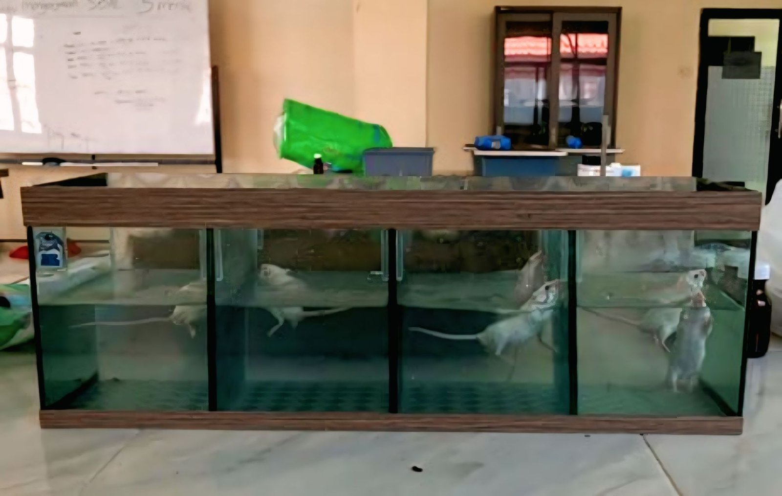
Tail Suspension Test (TST)
Mice were individually suspended by the tail using adhesive tape 1 cm from the tip and a horizontal bar 50 cm above the floor for 3 min daily over 7 consecutive days. In the present study, repeated TST exposure was employed as a sub-acute stress model to induce a transient stress-like state rather than as a validated model of chronic depression. Immobility time was defined as the duration during which the mice remained completely motionless and passively suspended (17). This approach was used as an exploratory paradigm to assess stress-related behavioral changes prior to pharmacological intervention. It is acknowledged that this method deviates from the conventional single-use TST protocol and may introduce adaptive or learned responses, which represents a limitation of the study.
Forced Swimming Test (FST)
Each mice was placed in a water-filled container for 6 min. The first 2 min were acclimatization and remaining 4 min were scored for immobility, defined as floating with minimal movements necessary for respiration (18). FST was conducted at T0 (baseline), T1 (after sub-acute stress exposure), and T2 (2 h after treatment administration) for behavioral screening purposes as presented in Figure 1. Repeated FST measurements were used for exploratory within subject comparison rather than as a validated longitudinal depression model. It is acknowledged that repeated exposure to FST may result in behavioral adaptation or learning effects, which could influence immobility time and represent a potential confounding factor (19).
Statistical Analysis
Data are presented as mean ± standard deviation (SD) and were analyzed using SPSS Statistics software. Data normality was assessed using the Shapiro–Wilk test, and homogeneity of variance was evaluated using Levene’s test (p > 0.05). Group comparisons were conducted using one-way analysis of variance (ANOVA), followed by Tukey’s post hoc test for multiple comparisons. A p-value (< 0.05) was considered statistically significant.
Results and Discussion
Characteristics and Phytochemical Screening of Extract:
C. alata leaf was extracted using the maceration method, yielding a thick, dark green extract with an aromatic odor and bitter taste. The yield % obtained was 12.389%. The % yield of the extracts is influenced by several factors, including the type of solvent used, extraction method, particle size of the sample (coarse or fine powder), presence of interfering compounds, sample to solvent ratio, temperature and extraction time (20).
| No. | Phytoconstituents | Result |
|---|---|---|
| 1 | Alkaloids | + |
| 2 | Flavonoids | + |
| 3 | Saponins | + |
| 4 | Terpenoids | - |
| 5 | Tannins | + |
| Notes: (+) indicates the presence of the active ingredient being tested. | ||
Preliminary phytochemical screening of the extract revealed the presence of flavonoids, alkaloids, saponins, and tannins, as summarized in Table 1. These classes of compounds are commonly reported in C. alata and are known to possess various biological activities. However, this screening remains qualitative and serves as an initial characterization of the extract composition. The negative result for terpenoids in the qualitative phytochemical screening may be related to the extraction solvent’s polarity, as ethanol typically favors the extraction of polar and semi-polar phytochemicals such as flavonoids and phenolic compounds. Solvent polarity has been shown to significantly influence the profile of secondary metabolites recovered from plant material (21). In addition, the concentration of terpenoids in the extract may be below the detection limit of the qualitative assay employed. The non-detection of terpenoids in the present screening does not prevent their presence but rather reflects the combined effects of solvent selectivity and assay sensitivity on phytochemical recovery (22).
LC-MS/MS Profiling
The chemical profile of the ethanolic extract of C. alata leaves was further investigated using LC–MS/MS analysis. The chromatogram of the extract showed multiple peaks across the retention time range, indicating the presence of diverse phytochemical constituents (Figure 2B–C). To ensure analytical reliability and exclude potential background interference, a blank chromatogram was analyzed under identical conditions. Comparison between the extract and blank chromatograms demonstrated that the major detected peaks originated from the sample rather than solvent or system-related artifacts (Figure 2A). Based on the retention time (RT), parent ion (m/z), and molecular weight, several major peaks were tentatively identified (Table 2). These identifications are predictive and tentative, as no authentic reference standards or MS/MS fragmentation library were employed to confirm the exact structures. The parent ions (precursor m/z) were used to search for chemical databases such as PubChem to obtain candidate compounds. Relative peak area (%) was used to describe the abundance of each detected compound in the extract, showing that flavonoid glycosides were the dominant class, followed by phenolic acids and fatty acids consistent with recent LC–MS/MS reports on C. alata and related extracts (23, 24). The presence of anthraquinone compounds including emodin has been reported in LC–MS analysis of C. alata extracts from various geographical sources (25). Notably, kaempferol 3-gentiobioside (24.3%) and luteolin (20.7%) were identified as the most abundant constituents, indicating that flavonoid derivatives represent the major fraction of the extract. These phytochemicals are well-aligned with the observed antidepressant-like activity, likely through combined antioxidant, neuroprotective, and neurotransmitter-modulating effects.
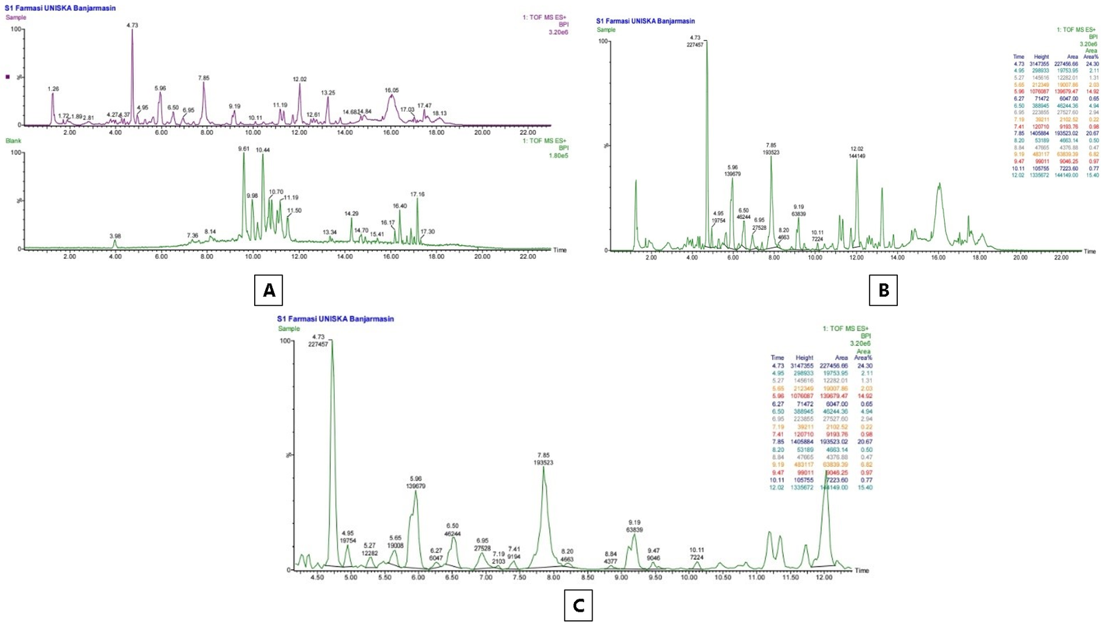
| No | Compound Name | Molecular Formula | RT (min) | Parrent (m/z) | Molecular Weight | Compound Composition (%) |
|---|---|---|---|---|---|---|
| 1 | Kaempferol 3-gentiobioside | C27H30O16 | 4.73 | 611.1608 | 610.52 | 24.3 |
| 2 | (3R)-6, 8-dihydroxy-3-[[2R, 5R, 6S)-5-hydroxy-6-methyloxan-2-yl] methyl]-3, 4-dihydroisochromen-1-one | C16H20O6 | 4.95 | 309.1448 | 308.327 | 2.1 |
| 3 | Kaempferol | C15H10O6 | 5.27 | 287.0558 | 286.24 | 1.3 |
| 4 | 7-methoxy-2, 2-dimethylchromene-6-carboxylic acid | C13H14O4 | 5.65 | 235.0976 | 234.25 | 2.0 |
| 5 | 4-[(2S)-3-hydroxy-2-methylpropyl]-2-methoxyphenol | C11H16O3 | 5.96 | 197.1181 | 196.24 | 14.9 |
| 6 | Luteolin 5, 3'-dimethyl ether | C17H14O6 | 6.27 | 315.0867 | 314.29 | 0.7 |
| 7 | (2Z, 4E)-5-(4-hydroxyphenyl) penta-2, 4-dienoic acid | C11H10O3 | 6.50 | 191.0711 | 190.20 | 4.9 |
| 8 | 7-hydroxyemodin | C15H10O6 | 6.95 | 287.0561 | 286.24 | 2.9 |
| 9 | 3, 3', 4, 5'-Tetrahydroxybibenzyl | C14H14O4 | 7.19 | 247.1337 | 246.26 | 0.2 |
| 10 | Emodin | C15H10O5 | 7.41 | 271.0615 | 270.24 | 1.0 |
| 11 | Luteolin | C15H10O6 | 7.85 | 287.0559 | 286.24 | 20.7 |
| 12 | Octyl glucoside | C14H28O6 | 8.2 | 293.2119 | 292.37 | 0.5 |
| 13 | 6'-O-Galloylsucrose | C19H26O15 | 8.84 | 495.1288 | 494.40 | 0.5 |
| 14 | 3, 4-dihydroxycinnamic acid (caffeic acid) | C9H8O4 | 9.19 | 181.1231 | 180.16 | 6, 8 |
| 15 | 1-(3, 5-dihydroxyphenyl) tridecan-2-yl acetate | C21H34O4 | 9.47 | 351.2535 | 350.49 | 1.0 |
| 16 | Eicosanedioic acid | C20H38O4 | 10.11 | 343.2950 | 342.51 | 0.8 |
| 17 | Octadecatetraenoic acid | C18H28O2 | 12.0 | 276.41 | 277.2173 | 15.4 |
| Note: Compound identification is tentative and based on RT, m/z, and molecular weight. Relative peak area (%) was calculated as the area of each peak divided by the total area of all detected peaks. | ||||||
However, the bioactivity in this study is only inferred based on compound presence and literature, as no pharmacokinetic or molecular assays were performed. The transition from chromatogram observation to compound listing allows for an overview of the chemical diversity in the extract and provides a tentative basis for future studies involving compound isolation, structure confirmation with standards, and mechanism validation.
It is important to note that RT and relative peak area are specific to the LC–MS/MS method used and may vary under different chromatographic conditions. Therefore, the identifications remain tentative and require further confirmation using authentic standards or MS/MS fragmentation library analysis for definitive structure assignments to ensure the highest level of analytical accuracy and prevent false positives.
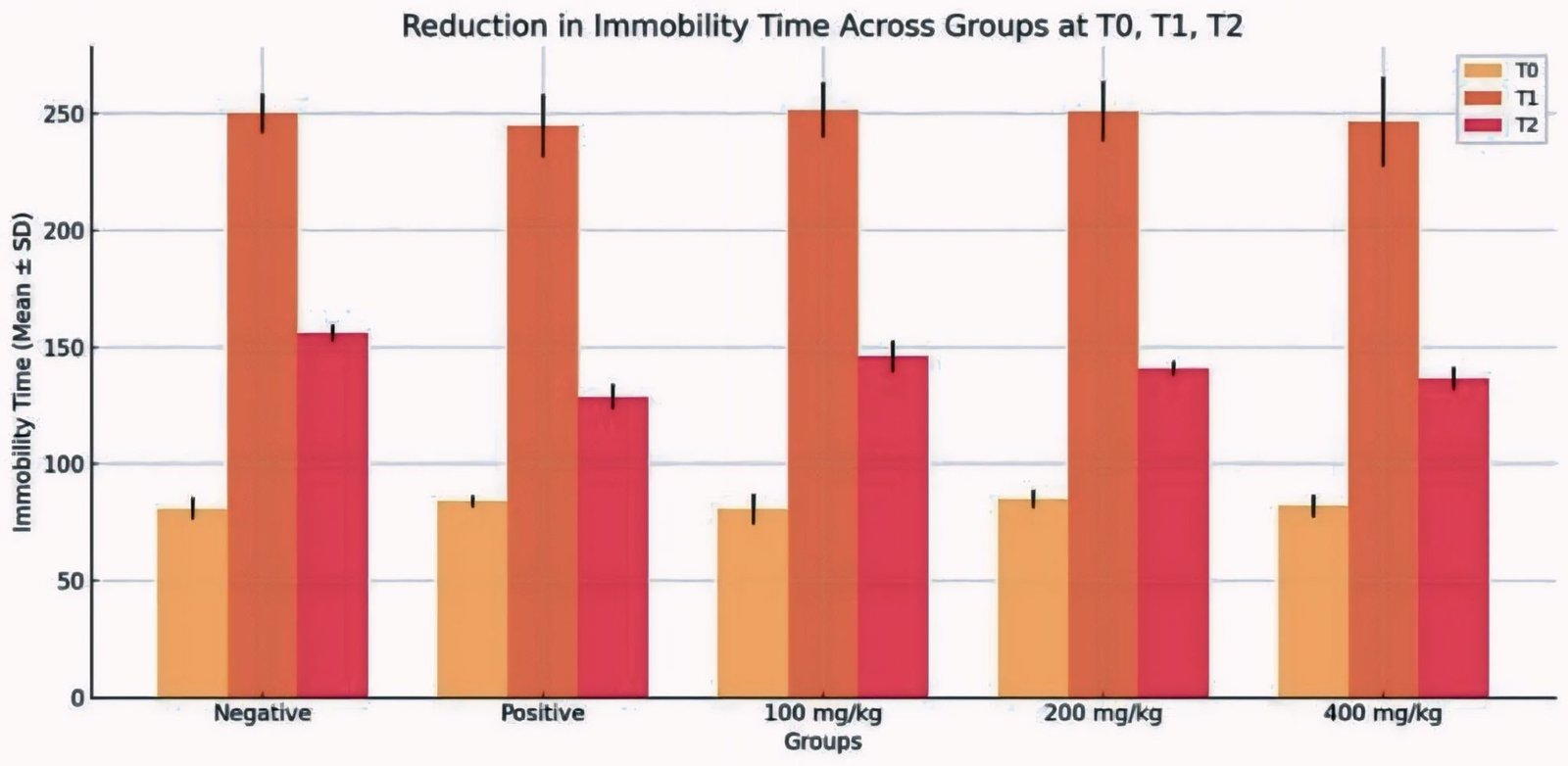
| Group | Mean SD | ||
|---|---|---|---|
| T0 | T1 | T2 | |
| Group I (Control) | 80.74 ± 4.53 | 250.12 ± 8.59 | 155.95 ± 3.78 |
| Group II (Standard) | 83.89 ± 2.59 | 244.66 ± 13.60 | 128.58 ± 5.40 |
| Group III | 80.58 ± 6.57 | 251.45 ± 11.90 | 145.90 ± 6.73 |
| Group IV | 84.80 ± 3.91 | 250.87 ± 2.93 | 140.65 ± 3.20 |
| Group V | 81.94 ± 4.93 | 246.24 ± 19.18 | 136.46 ± 4.86 |
Antidepressant Activity
The immobility time data are presented as mean ± standard deviation (SD), reflecting both the central tendency and the variability within each experimental group. The relatively low SD values across groups indicate consistent behavioral responses among animals within the same treatment group, supporting the reliability of the observed effects. The results of immobility time for each treatment group are summarized in Table 3. To provide a clearer visualization of the differences between groups, the Immobility Time presented in Figure 3.
The Forced Swimming Test (FST) was performed to assess antidepressant-like activity in mice. Data was collected at baseline (T0), after sub-acute stress exposure (T1), and after treatment (T2). Normality and Homogeneity of Variance were assessed before inferential analysis: Shapiro–Wilk test showed all groups were normally distributed (p > 0.05), and Levene’s test indicated homogeneity of variance (F = 1.067, p = 0.399). One-way ANOVA assesses differences among group means by partitioning the total variance into between-group and within-group components, expressed as sum of squares (SS) and degrees of freedom (df). The resulting F-statistics reflect the ratio of between-group variance to within-group variance. One-way ANOVA for T2 (immobility time) in Table 4 demonstrated a statistically significant difference among the groups (F (4, 20) = 21.465, p < 0.001).
| Sum of Squares | df | Mean Square | F | Siq. | |
|---|---|---|---|---|---|
| Between Groups | 2105.283 | 4 | 526.321 | 21.465 | 0.001 |
| Within Groups | 490.394 | 20 | 24.520 | - | - |
| Total | 2595.677 | 24 | - | - | - |
| Note: T2 represents immobility time measured after treatment. Data were analyzed using one-way ANOVA following confirmation of normal distribution (Shapiro–Wilk test, p > 0.05) and homogeneity of variances (Levene’s test, p = 0.399). | |||||
| Variable (I) | Variable (J) | Mean difference (I-J) | Sig. | Result |
|---|---|---|---|---|
| Negative | Positive | 27.36600* | 0.000 | Significant |
| Dose_1 | 10.05600* | 0.032 | Significant | |
| Dose_2 | 15.30000* | 0.001 | Significant | |
| Dose_3 | 19.49000* | 0.000 | Significant | |
| Positive | Negative | -27.36600* | 0.000 | Significant |
| Dose_1 | -17.31000* | 0.000 | Significant | |
| Dose_2 | -12.06600* | 0.008 | Significant | |
| Dose_3 | -7.87600 | 0.127 | Non-Significant | |
| Dose_1 | Negative | -10.05600* | 0.032 | Significant |
| Positive | 17.31000* | 0.000 | Significant | |
| Dose_2 | 5.24400 | 0.471 | Non-Significant | |
| Dose_3 | 9.43400* | 0.048 | Significant | |
| Dose_2 | Negative | -15.30000* | 0.001 | Significant |
| Positive | 12.06600* | 0.008 | Significant | |
| Dose_1 | -5.24400 | 0.471 | Non-Significant | |
| Dose_3 | 4.19000 | 0.672 | Non-Significant | |
| Dose_3 | Negative | -19.49000* | 0.000 | Significant |
| Positive | 7.87600 | 0.127 | Non-Significant | |
| Dose_1 | -9.43400* | 0.048 | Significant | |
| Dose_2 | -4.19000 | 0.672 | Non-Significant | |
| Note: Values represent mean differences between groups. * Indicates statistically significant differences at p < 0.05. Groups sharing no common superscript letters are significantly different based on Tukey’s post hoc test. Positive mean differences indicate higher immobility time in group I compared to group J. | ||||
Post hoc Tukey’s HSD analysis in Table 5 revealed dose-dependent effects the 400 mg/kg BW extract reduced immobility time significantly compared to the negative control (mean difference = -19.49, p < 0.001) and was not significantly different from the standard drug amitriptyline (mean difference = -7.876, p = 0.127). The 200 mg/kg BW dose exhibited intermediate activity, significantly different from the negative control (mean difference = -15.3, p = 0.001) but not from the 400 mg/kg BW group (p = 0.672). The 100 mg/kg BW dose showed moderate reduction, significant compared to negative control (mean difference = -10.056, p = 0.032) but less than higher doses. The negative control maintained high immobility time, confirming lack of antidepressant effect. These results indicate a clear dose-dependent reduction in immobility time, with the highest dose approaching the efficacy of the standard drug.
The observed antidepressant-like activity may be partly mediated by bioactive compounds identified in the extract via LC–MS/MS, including flavonol glycosides (kaempferol derivatives) (26), luteolin (27), phenolic acids (caffeic acid) (28), and anthraquinones (emodin) (29). Kaempferol derivatives (24.3%) and luteolin (20.7%) were identified as the dominant flavonoid constituents in the C. alata extract. These compounds have been widely reported to exhibit neuroprotective and antidepressant-like effects in recent preclinical studies and are known to cross the blood–brain barrier, enabling interaction with central nervous system targets (30).
Kaempferol and its glycoside derivatives have demonstrated antidepressant-like effects in recent studies through modulation of neuroinflammatory and intracellular signaling pathways. Recent evidence indicates that kaempferol can improve depression-like behaviors by suppressing neuroinflammation and regulating microglial activation, particularly through pathways such as PPARγ/STAT1 and inhibition of the NLRP3 inflammasome. These mechanisms are closely associated with the regulation of neuroinflammation, which is increasingly recognized as a key contributor to the pathophysiology of depression. In addition, kaempferol has been reported to influence neuronal survival and synaptic plasticity through its antioxidant and neuroprotective properties, further supporting its relevance in mood disorder modulation (31).
Luteolin has also been extensively investigated for its antidepressant-like properties in recent studies. Evidence suggests that luteolin can modulate monoaminergic neurotransmission by inhibiting serotonin (5-HT) reuptake and enhancing noradrenaline signaling, while also upregulating neurotrophic factors such as brain-derived neurotrophic factor (BDNF) and synaptic proteins involved in neuronal plasticity (32). Furthermore, luteolin has been shown to alleviate depression-like behavior in animal models by regulates oxidative stress, neuroinflammation, and neurotrophic signaling pathways in the hippocampus and prefrontal cortex, which are critical regions involved in mood regulation (33). Additional studies have also demonstrated that luteolin can modulate metabolic and autophagy-related pathways associated with depressive states, further supporting its multi-target neuropharmacological effects pathways (34). These findings highlight luteolin as a multifunctional flavonoid capable of influencing several biological processes relevant to depression.
Given their relatively high abundance in the extract, kaempferol derivatives and luteolin may contribute, at least in part, to the reduction in immobility time observed in the forced swimming test. Although previous reports have described the absence of observable toxicity of Cassia alata extracts at high doses in rodents (11), no toxicity or safety parameters were directly assessed in this study, and thus conclusions regarding safety cannot be definitively established. In addition, their antioxidant and anti-inflammatory properties may contribute to neuroprotective effects relevant to depressive-like states. However, it is important to emphasize that these mechanisms are inferred based on literature and compound presence, and were not directly evaluated in the present study.
Despite the observed behavioral effects, several limitations must be acknowledged. The study relied on a single behavioral assay (FST) as the primary endpoint without complementary models. Repeated exposure to both TST and FST may introduce behavioral adaptation or learning effects, potentially influencing immobility time. Moreover, no locomotor activity assessment (e. g., Open Field Test) was conducted; therefore, it cannot be excluded that reduced immobility may partially reflect increased general activity rather than a specific antidepressant-like effect. Furthermore, no biochemical or molecular analyses were performed to validate the proposed mechanisms, such as monoamine levels, monoamine oxidase activity, oxidative stress markers, or BDNF expression. The LC–MS/MS analysis provided only tentative compound identification based on mass data without confirmation using reference standards or fragmentation analysis. In addition, no dedicated toxicity or safety evaluation was conducted in this study; thus, conclusions regarding safety and tolerability remain limited.
Future studies should incorporate multiple behavioral paradigms, include locomotor control tests, apply repeated-measures statistical approaches, and perform molecular and pharmacokinetic investigations to better characterize the antidepressant potential and mechanism of action of C. alata extract. It is important to note that, although the Tail Suspension Test (TST) was applied repeatedly over 7 days, immobility data from TST were not systematically recorded and analyzed as an outcome measure in this study. Therefore, TST was primarily utilized as a sub-acute stress exposure rather than as an independent behavioral endpoint. This approach differs from conventional antidepressant screening protocols, where TST and FST are typically used as separate and complementary assays. The absence of longitudinal TST immobility data limits the interpretation of stress adaptation and behavioral progression over time. Future studies should include systematic recording and analysis of TST data alongside FST to provide a more comprehensive evaluation of antidepressant-like activity and stress-related behavioral responses.
Conclusion
This study demonstrates that the ethanolic extract of C. alata Leaves produced antidepressant-like behavioral effects in mice under the present experimental conditions, as indicated by a reduction in immobility time in the forced swimming test. Administration of the extract resulted in a dose-dependent decrease in immobility duration, with the highest tested dose (400 mg/kg body weight) showing the most pronounced behavioral effect. These findings are limited to acute behavioral observations and do not represent validated antidepressant efficacy or clinical relevance.
Although the extract was well tolerated during the experimental period, no dedicated toxicity or safety assessments were performed, and therefore conclusions regarding safety cannot be definitively established. Furthermore, the study is limited using a single behavioral paradigm and short-term evaluation, without molecular, biochemical, or repeated-measure analyses to support mechanistic interpretation. In conclusion, the ethanolic extract of C. alata Leaves exhibits preliminary antidepressant-like activity in an animal model. These findings provide an initial basis for further investigation; however, additional studies incorporating multiple behavioral paradigms, molecular and biochemical endpoints, long-term treatment designs, and comprehensive safety evaluations are required to substantiate the pharmacological relevance and translational potential of this plant extract.
Declarations
Acknowledgment
The authors would like to express their sincere gratitude to the Faculty of Pharmacy, Islamic University of Kalimantan Muhammad Arsyad Al-Banjari, for providing laboratory facilities and research support. The authors also gratefully acknowledge the Ministry of Education, Culture, Research, and Technology of the Republic of Indonesia, Directorate General of Higher Education, Research, and Technology, for the funding and support provided for this study.
Conflict of Interest
The authors declare no conflicting interest.
Data Availability
The data generated during and/or analyzed during the current study are available from the corresponding author on reasonable request.
Ethics Statement
All animal experiments were approved by the Komite Etik Penelitian Kesehatan Universitas Muhammadiyah Purwokerto (Approval No. KEPK/UMP/108/IX/2025) and conducted in accordance with relevant guidelines and regulations.
Funding Information
This work was supported by the Kementerian Pendidikan, Kebudayaan, Riset, Dan Teknologi, Direktorat Jenderal Pendidikan Tinggi, Riset, Dan Teknologi Republik Indonesia under Grant Number 2546/E2/DT.01.00/2024
References
- Tian YE, Xu M, Fang J, Wu Q, Zou X, Yan F, et al. Antidepressant-like active ingredients and their related mechanisms of functional foods or medicine and food homologous products. Digit Chin Med. 2023;6(1):9–27.
- Chen S, Tang Y, Gao Y, Nie K, Wang H, Su H, et al. Antidepressant potential of quercetin and its glycoside derivatives: a comprehensive review and update. Front Pharmacol. 2022;13:1–10.
- Friedrich MJ. Depression is the leading cause of disability around the world. JAMA. 2017;317(15):1517.
- Djohan SE, Lestari RD, Lestari E, Napitu IC. Emotional mental disorders and depression in adolescents. Healthc Nurs J. 2022;4(2):1–6.
- Tian H, Hu Z, Xu J, Wang C. The molecular pathophysiology of depression and the new therapeutics. MedComm (Beijing). 2022;3(3):e156.
- Rudra S, Faruque MO, Tahamina A, Emon NU, Haidar IK, Uddin SB. Neuropharmacological and antiproliferative activity of Tetrastigma leucostaphyllum. Saudi Pharm J. 2023;31(6):929–41.
- Gejalakshmi S, Harikrishnan N. Assessment of pharmacological potency of Clitoria ternatea. Res J Pharm Technol. 2025;18(10):4752–8.
- Sesa OE, Sulastry T, Muharram. Isolation and identification of secondary metabolite compounds from Cassia alata. Chemica. 2014;15(1):136–43.
- Sagnia B, Fedeli D, Casetti R, Montesano C, Falcioni G, Colizzi V. Antioxidant and anti-inflammatory activities of medicinal plants. PLoS One. 2014;9(8):1–10.
- Pamulaparthi A, Prathap VR, Banala M, Nanna RS. Experimental evaluation of antidepressant and antianxiety activities of Senna alata. Int J Curr Pharm Rev Res. 2016;8(4):60–3.
- Fatmawati S, Yuliana, Purnomo AS, Abu Bakar MF. Chemical constituents, usage and pharmacological activity of Cassia alata. Heliyon. 2020;6(7):1–11.
- Sofi NS, Yusransyah, Septiana D, Imam BS. Effectivity and evaluation of licorice root extract serum. Res J Pharm Technol. 2024;17(9):4142–8.
- Mittal S, Gupta P, Nigam V. Evaluation of antidepressant-like effect of Clitoria ternatea. Res J Pharm Technol. 2021;14(12):6437–41.
- Harborne JB. Phytochemical methods: a guide to modern techniques of plant analysis. 3rd ed. London: Chapman and Hall; 1998.
- Das A, Bharath M, Jeevanantham M, Kumar SM, Bindhu R. Phytochemical screening and antimicrobial activity of Syzygium cumini seed extract. Res J Pharm Technol. 2018;11(9):4096–100.
- Pratama RR, Sholikhah I, Sukardiman, Sahu RK, Widyowati R. Phytochemical compounds identification from Arcangelesia flava stems using LC-MS/MS. Pharmacogn J. 2023;15(4):528–34.
- Jodh R, Tawar M, Kachewar A. Evaluation of antidepressant activity of Tricholepis glaberrima using various paradigms. Res J Pharm Technol. 2022;15(12):5610–6.
- Mahmudah R, Hasanuddin S, Saleh A, Yuliastri WO, Isrul M. Antidepressant activity and identification of chemical compounds of mustard leaves extract. Res J Pharm Technol. 2019;12(7):3223–8.
- Patil S, Nargatti P, Shikalgar T, Naikwade N. Evaluation of antidepressant activity of Ficus carica leaves extract. Res J Pharm Technol. 2021;14(3):1267–73.
- Tsaturyan A, Sahakyan L, Hayrapetyan L, Minasyan E, Chakhoyan A, Mirzoyan V, et al. Effect of solvent polarity on extraction of bioactive compounds. Pharmacia. 2025;72:1–9.
- Azzahra VO, Mardiana S, Suharyadi S, Sianipar RJ, Ramadhan DS. Effect of solvent polarity on extraction yield and phytochemical composition. Biol Med Nat Prod Chem. 2025;14(2):1273–84.
- Saptarini NM, Mustarichie R, Hasanuddin S, Corpuz MJAT. Cassia alata antifungal activity and molecular docking study. Pharmaceuticals. 2024;17(3):380.
- Colin M, Claudiana NS, Kaffah A, Hasanah A, Megantara S. Review on Cassia alata bioactive compounds. Drug Des Devel Ther. 2024;18:4427–47.
- Angelina M, Mardhiyah A, Dewi RT, Fajriah S, Muthiah N, Ekapratiwi Y, et al. Physicochemical and phytochemical standardization of Cassia alata leaves. Pharmacia. 2021;68(4):947–56.
- Ishisaka M, Kakefuda K, Yamauchi M, Tsuruma K, Shimazawa M, Tsuruta A, et al. Luteolin shows an antidepressant-like effect. Biol Pharm Bull. 2011;34(9):1481–6.
- Pena JB, Kim CA, Lee HL, Yoon SY, Kim HJ, Hong EY, et al. Luteolin mediates antidepressant-like effects via GABAA receptor modulation. Arch Pharm Res. 2014;37(2):263–9.
- Kandilarov IK, Zlatanova HI, Georgieva-Kotetarova MT, Kostadinova II, Katsarova MN, Dimitrova SZ, et al. Antidepressant effect of plant extract combinations. Folia Med (Plovdiv). 2018;60(1):110–6.
- Zeng P, Wang XM, Ye CY, Su HF, Fang YY, Zhang T, et al. Mechanistic insights into the antidepressant effect of emodin. Aging (Albany NY). 2021;13(11):15078–99.
- Wang X, Zhang W, Wang H, Zhao Y, Wang P, Wang R, et al. Dietary kaempferol attenuates cognitive decline. Food Funct. 2026;17(1):588–603.
- Su P, Liu L, Gong Y, Peng S, Yan X, Bai M, et al. Kaempferol improves depression-like behaviors. Int Immunopharmacol. 2024;143:1–10.
- Zhou J, Wu Z, Zhao P. Luteolin and its antidepressant properties. J Pharm Anal. 2025;15(4):1–10.
- Wu X, Xu H, Zeng N, Li H, Yao G, Liu K, et al. Luteolin alleviates depression-like behavior. CNS Neurosci Ther. 2024;30(3):1–10.
- Mokhtari T, Lu M, El-Kenawy AEM. Potential anxiolytic and antidepressant-like effects of luteolin. Int Immunopharmacol. 2023;122:1–10.