RESEARCH ARTICLE
Antiprostatic Activities of Hippocratea africana Root Extract and Fractions Against Testosterone-Induced Benign Prostatic Hyperplasia in Rats
Sciences of Pharmacy|Vol. 5, Issue 2, pp. 210-223 (2026)
CC BY 4.0-2026 Authors
Views
Downloads
Shares
Received
Feb 26, 2026Revised
May 5, 2026Accepted
Jun 3, 2026Published
Jun 8, 2026
Abstract
Hippocratea africana (Willd. ) Loes. ex Engl. (Celastraceae) syn. Loeseneriella africana (Willd. ) N. Hallé root which is used as medicine traditionally to treat various diseases by the Ibibios was investigated for antiprostatic effect against testosterone-induced benign prostatic hyperplasia (BPH). The dried root powder was cold extracted in 50% ethanol and the extract dissolved in distilled water and partitioned with dichloromethane (DCM) to obtain DCM and aqueous fractions of H. africana root. BPH was induced in groups of male rats (n=5) using testosterone propionate (3 mg/kg). Based on previously determined median lethal dose, the root extract and fractions of H. africana (200-600 mg/kg) were investigated for effects on prostate weight and histology, prostate sensitive antigen (PSA), testosterone levels, lipid profile, oxidative stress markers, semen analysis and testis histology of rats with testosterone propionate induced BPH. Finasteride (5 mg/kg) was used as standard drug. The root extract/fractions were found to cause significant (p < 0.05 - 0.01) decreases in PSA and testosterone levels, reductions in prostate and testis weights, improvement of semen volume and health, elevations of antioxidant enzymes (SOD, CAT, GPX) and molecule (GSH) and also reduced MDA level. These results suggest that the root extract and fractions of H. africana possess antiprostatic potentials against testosterone-induced BPH in rats which may be due to the antioxidant activities of its phytochemical constituents.
Introduction
The malignant proliferation of stromal and epithelial cells resulting in the enlargement of prostate gland is a medical condition known as benign prostatic hyperplasia (BPH), which may or may not be associated with lower urinary tract symptoms (1). BPH occurs more in men of age 51 and above and its prevalence rate increases with age (2). Statistics have shown an upsurge in global burden of BPH and further revealing that more than half of the men at age 60 have BPH while age 85 and above have a prevalence rate of 90% (3). Oxidative stress, which occurs as a result of imbalance between the free radicals in biological systems and the endogenous antioxidants, has been associated with the development and progression of BPH (4). Free radicals generated from various physiological and metabolic processes in the body result in oxidative stress, and cause damages to fatty tissues, DNA, proteins and cell membrane resulting in inflammation and organ dysfunction (5). Other factors such as inflammatory mediators, growth factors, sex hormones, dietary factors, neurotransmitters and environmental factors also play key roles in the pathogenesis of BPH (6).
Plants have been a reservoir for therapeutic agents used to sustain the primary health care needs of man globally especially in developing countries. In Nigeria, a vast majority of people, irrespective of societal status, still patronise herbal medicine for their health care needs due to its accessibility and low cost (7). There are claims of efficacy of some medicinal plants used locally to treat prostate disorders, one of which is Hippocratea africana.
H. africana (Willd. ) Loes. ex Engl. (Celastraceae) syn. Loeseneriella africana (Willd. ) N. Hallé is a perennial tropical plant that is found widely in tropical African rainforests (8). It is also called the African paddle-pod and 'Eba enang enang' by the Ibibios of Nigeria. The plant's roots have been widely employed in ethnomedicine by the Ibibios of the southern part of Nigeria to treat many ailments such as fever, convulsion, malaria, ulcers, body pains, infections, diabetes, and diarrhea, among others (9). The plant's root is also used for its antidotal or anti-poison potential to treat liver diseases such as jaundice and hepatitis (10-12). Previous scientific reports have shown that the root extract possesses antimalarial (9, 13), angioedema and antinociceptive (14), antidiabetic and hypolipidemic (15, 16), antidiarrhoeal and antiulcer (17), hepatoprotective (18-21), antileishmanial, cytotoxicity and cellular antioxidant (22), antibacterial, anticonvulsant and depressant (23), in vivo alpha-amylase and alpha-glucosidase inhibitory (24), in vitro antioxidant (17, 25), renoprotective (26-28), genotoxic (29), testiculoprotective (30) and cardioprotective (31) activities. GCMS analysis of the root fractions revealed the presence of spiro hexane-1-carboxylic acid, ethyl ester, 3-methoxy-2-methylphenol, 2, 3-benzofuran dione, 6-hydroxy-4-(p-hydroxy benzyl), ᵟ-3-Carene and α-terpineol in ethyl acetate fraction (32), monoterpenes (thujene, limonene, linalool, α-phellandrene, α-terpineol, and sabinene) and sesquiterpenes (dehydromevalonic lactone), in the n-hexane fraction of the root extract (18). Also, two xanthones, 1, 3, 6, 7-tetrahydroxyxanthone and 1, 3, 6-trihydroxy-7-methoxyxanthone have been isolated from the root of H. africana (25). The root of H. africana is used traditionally in Ibibio traditional medicine to treat prostate disorders. Although there are claims of its efficacy locally, there is no documented evidence or scientific proof to these claims. We report the activities of root extract and fractions of H. africana against testosterone-induced benign prostatic hyperplasia in rats.
Materials and Methods
Plants collection and extraction
In July 2025, fresh roots of H. africana were collected in bushes in the Uruan area, Akwa Ibom State, Nigeria. Prof Magaret Bassey, a taxonomist from the Department of Botany and Ecological Studies, University of Uyo, Uyo, Nigeria, identified and authenticated the plant. Herbarium specimen UUDPHB 30 (i) was deposited at the Department of Pharmacognosy and Natural Medicine Herbarium, University of Uyo. The fresh roots of H. africana were washed, reduced into smaller pieces, and shade-dried in the laboratory for two weeks. These were later pulverized using an electric grinder. The pulverized root of H. africana (1 kg) was soaked in ethanol (Sigma-Aldrich, USA), (50%) for 72 h. The liquid filtrate obtained was concentrated in a rotary evaporator at 40˚C. The crude extract was dissolved in 500 mL of distilled water, amounting to 20 g, and partitioned with dichloromethane (DCM (Sigma-Aldrich, USA), 5 x 500 mL) on an equal volume till no color change was observed to obtain DCM and aqueous fractions. The extract and fractions were stored in a refrigerator at 4˚C until used for the experiment.
Experimental animals
In this study, male albino Wistar rats (125 – 140 g) sourced from the University of Uyo Animal House and sheltered in plastic cages were used. The rats were fed on standard pelleted diet and water ad libitum, kept under ambient temperature (28 ± 2 °C) and illuminated environment of 12:12 h dark/light cycle. The animals were allowed to acclimatize to laboratory conditions for two weeks prior to the commencement of the experimental procedures. The animal study was approved by the College Health Sciences Animal Ethics Committee, University of Uyo (UU/CHS/IHREC/25/VOL.1/14).
Induction of Benign Prostactic Hyperplasia (BPH) in The Rats
Benign prostactic hyperplasia (BPH) was induced in rats through daily intraperitoneal injections (i. p) of TP (3 mg/kg, dissolved in corn oil) for 28 days (33).
Study Design
The rats were randomized into eight groups of five animals each. Group 1 (control) received distilled water. Group 2 (BPH-induced control) received a testosterone propionate (TP) injection along with distilled water. Group 3 was treated with the standard drug, finasteride (5 mg/kg), and a TP injection. Based on the previously established LD50 value of 2449.49 mg/kg [9], Group 4 (low dose) was administered 200 mg/kg of H. africana root extract and a TP injection. Groups 5 and 6 were treated with 400 mg/kg and 600 mg/kg of the H. africana root extract, respectively, alongside a TP injection. Groups 7 and 8 were treated with 400 mg/kg of the aqueous and dichloromethane fractions of the H. africana root extract, respectively, concomitantly with a TP injection.
Rat body weights were recorded at the beginning and at the end of the experiment. All test materials (H. africana root extract and fractions) were administered daily for 28 days. Following the final administration and an overnight fast, the rats were anesthetized via intraperitoneal injection of a ketamine and xylazine mixture (0.1 mL/100 g body weight), containing 90 mg/kg ketamine and 9 mg/kg xylazine. Blood samples were collected by cardiac puncture into plain centrifuge tubes, left to clot for 15 min, and centrifuged at 2500 rpm for 15 min to separate the serum for biochemical assays. Subsequently, liver and prostate tissues were immediately dissected, weighed, and fixed in 10% formaldehyde for histopathological analysis. The prostate index was calculated as the ratio of prostate weight to total body weight.
Measurement of Serum Testosterone and PSA Levels
The serum levels of testosterone and PSA were assayed using ELISA (Cayman, USA and Novatein Biosciences, Woburn, MA, USA, respectively) following the manufacturer’s instructions.
Evaluation of Progressive Motility, Viability, Count, and the Structural Abnormality of Sperm
The caudal piece of epididymis was isolated to retrieve the sperm samples. Initially, the epididymal part was finely minced in 5 mL of physiological-saline and was incubated for 30 min at 37 °C for spermatozoa releasing of the epididymal ducts. Sperm progressive motility % was noted through the phase-contrast microscope at 400x (34). Sperm viability was assessed, by eosin or nigrosin staining, accompanied by microscopic evaluation. Moreover, a hemocytometer was employed to count epididymal sperm in the suspension (35). Furthermore, morphological anomalies of head, tail, and mid piece of sperm were determined in % using the method of Filler (36). The apparent abnormal characteristics included (i) the size and shape of spermatozoa heads (bigor small heads) with lighter and emphasized curvature; (ii) intermediary pieces’ defects that result in untied heads; and (iii) defects of tails (short, multiple, folded, and broken tails). Photomicrograhs of the semen samples were taken under the microscope to assess the spermatozoa population at x100.
Assessment of Lipid Profile
Serum cholesterol, triglyceride and high-density lipoprotein (HDL) levels of the treated rats were measured using standard colorimetric methods (37). These determinations were done spectrophotometrically using Fortress Diagnostic Kits® according to standard procedures of manufacturer’s protocols. Low and very low-density lipoprotein (LDL and VLDL) were estimated from the formula of Friedwald et al. (38).
Antioxidant Enzymes (Oxidative Stress Markers) Estimation
The sera samples collected from the rats were used for the determination of malondialdehyde (MDA) content (39), superoxide dismutase (SOD) (40), catalase (CAT) (41), glutathione peroxidase (GPx) (42) and reduced glutathione (GSH) (43).
Histopathological Studies
The testes and prostates of the animals were surgically removed, weighed and fixed in 10% formaldehyde for histological processes. According to Haematoxylin and Eosin method (44), the organs were carefully dissected out, trimmed of all fat and blotted dry to remove any blood. They were then fixed in 10 % formal saline (fixation). The fixed tissues were transferred to a graded series of ethanol (Dehydration). On day 1, the tissues were placed in 70% alcohol for 7 h, and then be transferred to 90% alcohol and left in the latter overnight. On day 2, the tissues were passed through three changes of absolute alcohol for an h each then cleared in xylene (clearing). Once cleared, the tissues were infiltrated in molten paraffin wax in the oven at 58 °C. Three changes of molten paraffin wax (impregnation) at one-h intervals were made, after which the tissues were embedded (embedding) in wax and blocked out. Serial vertical sections of 5µm thick were obtained from a solid block of tissue (microtomy) fixed on clean albuminized slides to prevent sections from pulling off the slides and later stained with haematoxylin and eosin staining techniques, after which they were passed through grades of alcohol, cleared in xylene and mounted in DPX (Distyrene - Plasticizer and xylene) mountant and observed under digital light microscope at Department of Chemical Pathology, University of Uyo Teaching Hospital, Uyo. Morphological changes were observed; recorded and Histologic photomicrographs were taken and interpreted by Histopathologist.
Statistical Analysis
Data obtained from this work were analysed statistically using Graphpad Instat, (California, USA). ANOVA (one –way) followed by a post-test (Tukey-Kramer multiple comparison test) were used. Differences between means were considered significant at 5% level of significance i. e., p ≤ 0.05.
Results
Yields of Crude Extract and Solvent Fractions
The % yields of the crude extract and solvent fractions were crude 26.02%, DCM 5.01%, and Aqueous 93.44%.
Effect on Organ Weights
The effect of administration of root extract and fractions of H. africana on testes and prostate weights of rats with testosterone-induced BPH is as shown in Table 1. The prostate weights of rats were significantly (p < 0.01) increased following testosterone (3 mg/kg/day) administration for 28 days relative to normal control. Concomitant administration of root extract and fractions of H. africana and testosterone to male rats, did not change prostate weights of rats significantly (p > 0.05) relative to the control except in the groups treated with finasteride (standard drug) and 400 mg/kg of the extract where significant (p < 0.01) reduction in prostate weights relative to testosterone-only treated group was observed. Similarly, testosterone only treated group was observed to have elevated prostate index value relative to the normal control, and all the extract/fractions treated groups. Groups of rats treated with DCM fraction, finasteride and extract (400 mg/kg) recorded the lowest prostate index values. The testes weight of testosterone only treated group was observed to be significantly (p < 0.05) increased relative to normal control, while finasteride, extract and fractions produced non dose-dependent but significant (p < 0.05-0.01) reduction in testes weights of rats relative to testosterone only treated group.
| Parameters/ Treatments | Dose mg/kg | Testes (mg) | Prostate (mg) | Body weight (g) | Prostate index |
|---|---|---|---|---|---|
| Normal Control | - | 1.56 ± 0.21 | 0.48 ± 0.02 | 228.5 ± 6.22 | 0.0021 |
| Testosterone Only | 3 | 2.97 ± 0.19b | 0.76 ± 0.05b | 240.0 ± 20.70 | 0.0031 |
| Finasteride+TTT | 5 | 1.44 ± 0.11d | 0.60 ± 0.03d | 224.0 ± 9.12 | 0.0026 |
| Extract+TTT | 200 | 1.51 ±0.06d | 0.71 ± 0.10 | 230.75 ± 11.04 | 0.0030 |
| 400 | 1.59 ±0.76d | 0.61 ± 0.04d | 231.75 ± 7.56 | 0.0026 | |
| 600 | 1.76 ±0.07d | 0.68 ± 0.03a | 221.66 ± 7.53 | 0.0030 | |
| Aqueous Fraction+TTT | 400 | 1.38 ± 0.22f | 0.63 ± 0.05 | 228.75 ± 9.62 | 0.0027 |
| Dichloromethane Fraction+TTT | 400 | 1.61 ± 0.06d | 0.64 ± 0.09 | 252.0 ± 8.84 | 0.0025 |
| Note: Data are expressed as MEAN ± SEM, Significant at ap < 0.05, bp < 0.01 when compared to control; Significant at dp < 0.05, fp < 0.001 when compared to testosterone only control (n=5). | |||||
Effect of Root Extract and Fractions of H. africana on Lipid Profile of Rats with Testosterone-Induced BPH
Administration of testosterone propionate (3 mg/kg/day) was observed to caused elevations of lipid profile parameters such as total cholesterol, triglyceride, low density lipoprotein and very low density lipoprotein significantly (p < 0.05-0.001) and reduction in high density lipoprotein level relative to control. However, decreases in the levels of total cholesterol, triglyceride, high density lipoprotein, low density lipoprotein and very low density lipoprotein were observed following concomitant treatment of the rats with root extract / fractions of H. africana (200-600 mg/kg) and testosterone propionate (3 mg/kg i. p) for 28 days. These decreases were significant (p < 0.05-0.001) especially in the groups treated with the low and high doses of the extract (200 and 600 mg/kg) as well as the fractions treated groups (aqueous and DCM) when compared to testosterone only group. Similarly, finasteride caused reductions in the lipid parameters levels which were significant (p < 0.05-0.001) in total cholesterol and low density cholesterol levels when compared to testosterone only group (Table 2).
| Treatment | Dose mg/kg | Total cholesterol (mMol/L) | Triglyceride (mMol/L) | HDL-C (mMol/L) | LDL-C (mMol/L) | VLDL (mMol/L) |
|---|---|---|---|---|---|---|
| Normal Control | 10 mg/mL | 2.96 ± 0.08 | 1.21 ± 0.02 | 1.54 ± 0.04 | 0.21 ± 0.04 | 0.18 ± 0.02 |
| Testosterone only | 3 | 4.23 ± 0.26b | 1.45 ± 0.05 | 1.32 ± 0.03 | 0.48 ± 0.02b | 0.35 ± 0.01b |
| Finasteride +TTT | 5 | 2.96 ± 0.03e | 1.24 ± 0.05 | 1.36 ± 0.06 | 0.27 ± 0.01f | 0.26 ± 0.02 |
| Crude extract + TTT | 200 | 2.50 ± 0.11f | 0.99 ± 0.07d | 1.21 ± 0.05a | 0.32 ± 0.03d | 0.27 ± 0.02 |
| 400 | 3.56 ± 0.23 | 1.30 ± 0.07 | 1.44 ± 0.08 | 0.22 ± 0.02f | 0.12 ± 0.01f | |
| 600 | 2.80 ± 0.20f | 1.06 ± 0.07d | 1.20 ± 0.05b | 0.16 ± 0.02f | 0.14 ± 0.03f | |
| Aqueous fraction+ TTT | 400 | 2.43 ± 0.12f | 0.94 ± 0.05d | 1.09 ± 0.03c | 0.30 ± 0.05e | 0.15 ± 0.04f |
| Dichloromethane fraction+TTT | 400 | 2.80 ± 0.17f | 1.10 ± 0.07 | 1.24 ± 0.05a | 0.18 ± 0.02f | 0.15 ± 0.01f |
| Note: Data are expressed as MEAN ± SEM, Significant at ap < 0.05, bp < 0.01, cp < 0.001, when compared to control; Significant at dp < 0.05, ep < 0.01, fp < 0.001 when compared to testosterone only control (n=5). | ||||||
Effect of H. africana Root Extract and Fractions on Oxidative Stress Markers in Rats with Testosterone-Induced BPH
The effect of administration of root extract and fractions of H. africana on oxidative stress markers of rats with testosterone-induced BPH is as shown in Table 3. Treatment of rats with testosterone (3 mg/kg) daily for 28 days was found to decrease significantly (p< 0.05-0.01) the levels of oxidative stress markers (CAT, SOD, GSH, GPx) and also increased significantly (p < 0.05) the MDA level of rats when compared to normal control. However, concomitant administration of root extract and fractions of H. africana with testosterone was found to mitigate these effects and caused marked elevations of the enzymatic and non-enzymatic endogenous antioxidants in the treated rats groups when compared to the testosterone only groups. These elevations were not dose-dependent but were significant (p < 0.05-0.001) in all treatment groups. MDA levels were only reduced significantly (p < 0.05) in groups treated with middle dose (400 mg/kg), dichloromethane fraction and finasteride when compared to testosterone-only treated group.
| Treatment | Dose (mg/kg) | SOD (U/mL) | CAT (U/g of protein) | GPx (µg/mL) | GSH (µg/mL) | MDA (µMol/mL) |
|---|---|---|---|---|---|---|
| Normal Control | - | 0.36 ± 0.02 | 4.66 ± 0.45 | 0.055 ± 0.002 | 2.46 ± 0.11 | 0.26 ± 0.01 |
| Testosterone only | 3 | 0.17 ± 0.01c | 0.46 ± 0.01a | 0.040 ± 0.00a | 0.31 ± 0.01b | 0.42 ± 0.01c |
| Finasteride +TTT | 5 | 0.30 ± 0.02f | 2.79 ± 0.56f | 0.044 ± 0.004 | 1.97 ± 0.21e | 0.22 ± 0.01f |
| Extract+TTT | 200 | 0.38 ± 0.03f | 2.69 ± 0.31f | 0.048 ± 0.002 | 2.20 ± 0.10f | 0.38 ± 0.05 |
| 400 | 0.43 ± 0.02f | 3.39 ± 0.61f | 0.052 ± 0.005d | 2.72 ± 0.16f | 0.30 ± 0.01d | |
| 600 | 0.32 ± 0.01f | 3.84 ± 0.25f | 0.059 ± 0.001e | 2.63 ± 0.01f | 0.39 ± 0.02 | |
| Aqueous Fraction +TTT | 400 | 0.31 ± 0.02f | 2.98 ± 0.06f | 0.045 ± 0.003 | 2.08 ± 0.13f | 0.35 ± 0.02 |
| DCM fraction +TTT | 400 | 0.31 ± 0.01f | 2.37 ± 0.06f | 0.052 ± 0.001d | 2.37 ± 0.01f | 0.26 ± 0.02e |
| Note: Data are expressed as MEAN ± SEM, Significant at ap < 0.05, bp < 0.01, cp < 0.001, when compared to control; Significant at dp < 0.05, ep < 0.01, fp < 0.001 when compared to testosterone only control (n=5). | ||||||
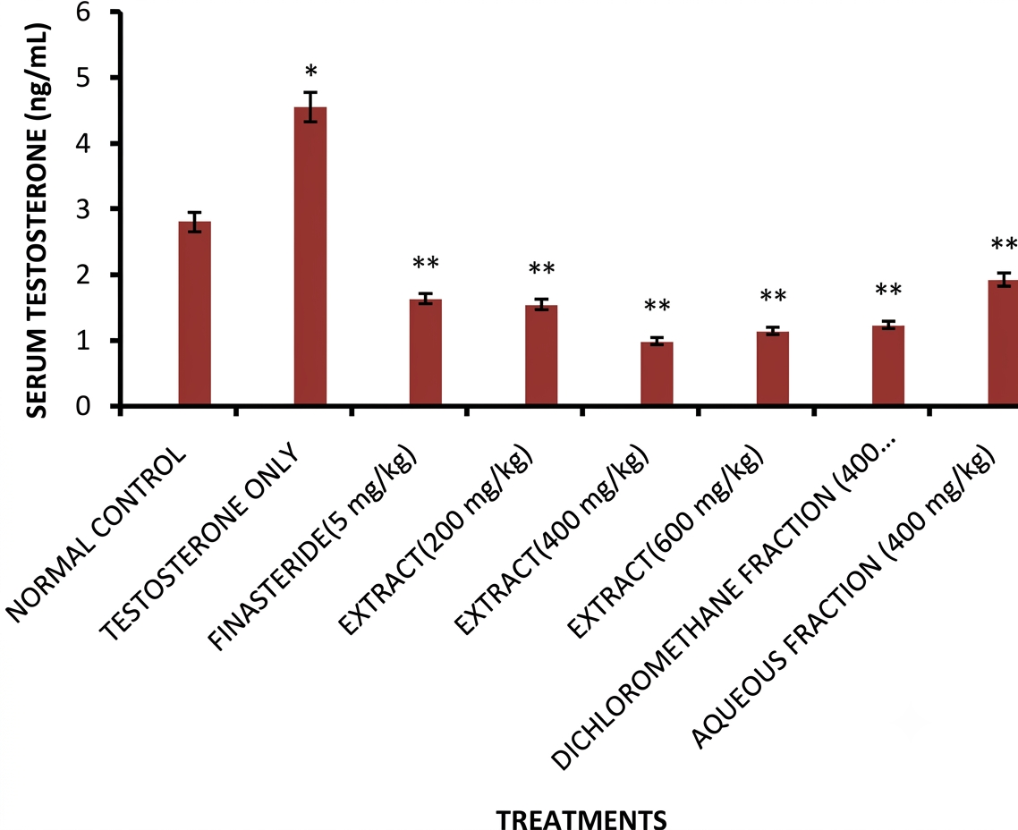
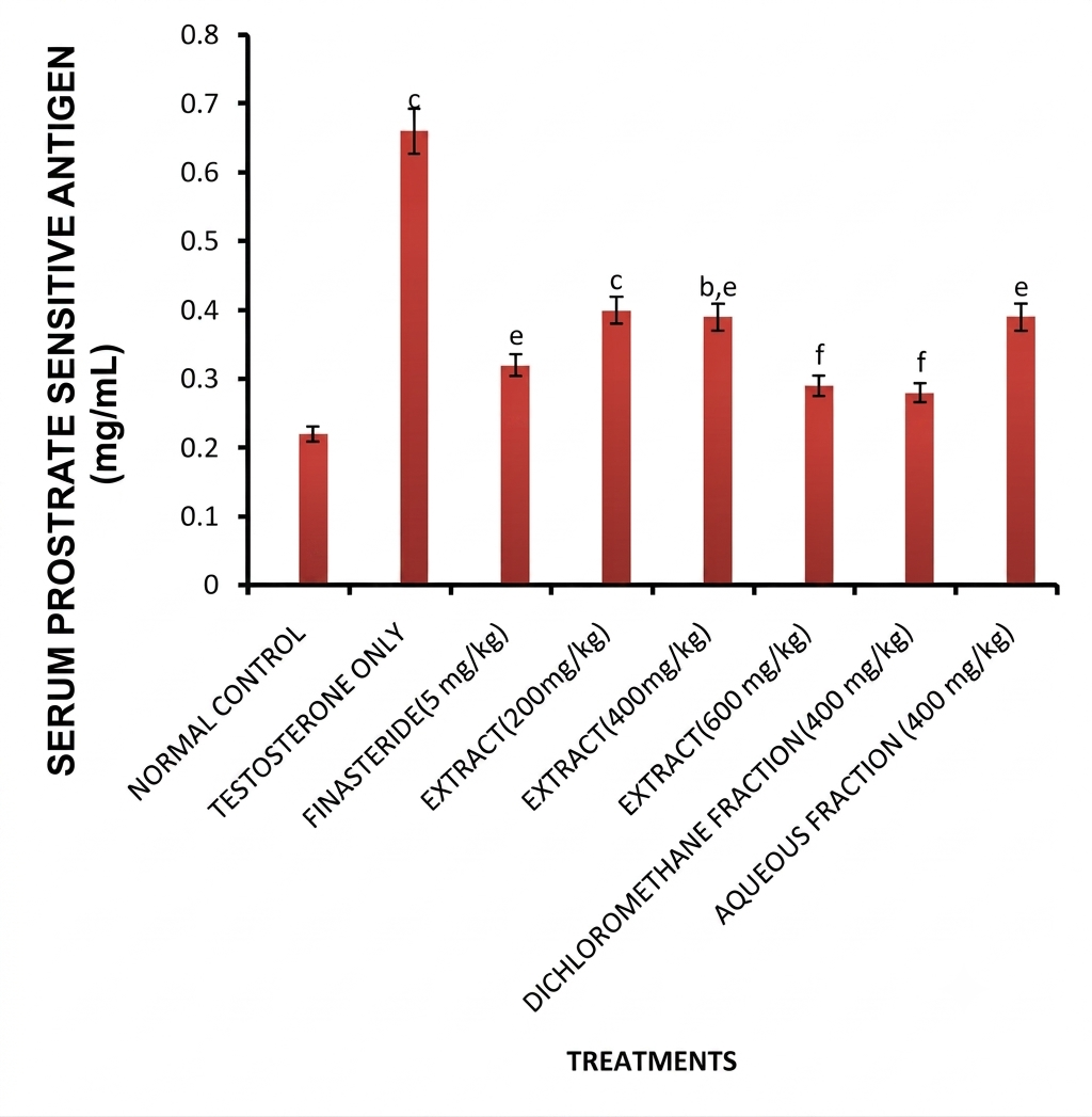
Effect of H. africana Root Extract on Serum Testosterone and Prostate-Specific Antigen (PSA) Levels in Rats with Testosterone-Induced BPH
The effect of administration of root extract and fractions of H. africana on serum testosterone and Prostate sensitive Antigen (PSA) levels of rats with testosterone-induced benign prostatic hyperplasia are as shown in Figures 1 and 2. Treatment of rats with testosterone (3 mg/kg i.p) daily for 28 days caused significant (p < 0.01-0.001) elevations of serum testosterone and prostate sensitive antigen levels when compared to normal control. The concurrent administration of root extract and fractions of H. africana to male rats caused reductions in testosterone and prostate sensitive antigen levels which were significant (p < 0.01-0.001) in groups treated with 200, 400 and 600 mg/kg of extract, finasteride, aqueous and dichloromethane fractions when compared to testosterone-only treated group. Aqueous fraction-treated group was found to produce the most significant (p < 0.001) reductions in testosterone and PSA levels when compared to testosterone only treated group.
Effect of H. africana Root Extract and Fractions on Sperm Parameters in Rats with Testosterone-Induced BPH
Table 4 shows the seminal analysis of semen from rats with testosterone-induced BPH. The semen in all treatment group was found to be milky in appearance, while the semen volume in the group administered with testosterone alone was found to be small (0.01 mL). There was a marked improvement in the semen volume of rats treated with the root extract and fractions (0.02 -0.04 mL) with the DCM fraction treated group having the highest volume (0.043 mL). The PH of the semen samples from all the groups was 8.0. The extract/fraction-treated groups were found to have a higher % of viable sperm cells (78-90%) compared to the testosterone only-treated group (55%). Finasteride treated group had 90% viable cells while control had 80%. Viscosity of semen in all the groups was normal. The average number of cells was 33.25 x 10⁶ in the testosterone only treated group, but non-dose dependent improvements were observed in extract / fractions treated groups with high dose (600 mg/kg) having 130.75 x 10⁶ cells, while DCM fraction treated group recorded average cell of 126.75 x 10⁶ cells and finasteride group 91.0 x 10⁶ cells. The % of active sperm cells in the extract-treated groups ranged from 55-80% non-dose-dependent compared to 50% recorded in the testosterone only group. The % of dead sperm cells in the testosterone only group was found to be about 10% compared to 5% in the middle dose (400 mg/kg) and high dose (600 mg/kg) treated-groups as well as the groups treated with the various fractions. The standard drug (finasteride) group had 5 % dead sperm cell.
| Parameters | Normal Control | Testosterone only | Finasteride | Extract 200 mg/kg | Extract 400 mg/kg | Extract 600 mg/kg | Aqueous fraction | DCM fraction |
|---|---|---|---|---|---|---|---|---|
| App | Milky | Milky | Milky | Milky | Milky | Milky | Milky | Milky |
| Volume | 0.03 mL | 0.01 mL | 0.02 mL | 0.02 mL | 0.03 mL | 0.03 mL | 0.03 mL | 0.043 mL |
| PH | 8.0 | 8.0 | 8.0 | 8.0 | 8.0 | 8.0 | 8.0 | 8.0 |
| Viability | 80% | 55% | 90% | 78% | 80% | 90% | 90% | 90% |
| Viscosity | Normal | Normal | Normal | Normal | Normal | Normal | Normal | Normal |
| Average cells (cell x106) | 85.25 | 33.25 | 91.00 | 104.00 | 78.75 | 130.75 | 97.00 | 126.75 |
| Normal | 70% | 65% | 75% | 70% | 75% | 65% | 70% | 70% |
| Abnormal | 30% | 35% | 25% | 30% | 25% | 35% | 30% | 30% |
| Active | 90% | 50% | 70% | 60% | 70% | 55% | 60% | 80% |
| Sluggish | 5% | 40% | 25% | 30% | 25% | 40% | 35% | 15% |
| Dead | 5% | 10% | 5% | 10% | 5% | 5% | 5% | 5% |
| Sperm | 500 | 200 | 600 | 400 | 450 | 600 | 600 | 600 |
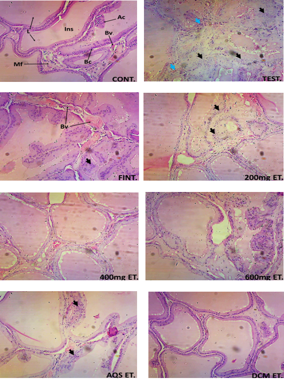
Effect of root extract and fractions of H. africana on histology of rats prostate with testosterone induced benign prostatic hyperplasia Histological sections of prostate of rats receiving various treatments at magnification (x100) stained with H& E method revealed that Group 1 (normal control, CONT) treated distilled water (10 mL/kg) had prostate tissue demonstrating a normal histo-architecture of a prostate gland, with well presented glandular lumen, acini epithelial cells, intraluminal secretions, lined basement cells, well structured fibrocyte nuclie, smooth muscle fibre and interluminal blood vessel within the prostatic stroma, no evidence of pathology was seen (Figure 1). The group (Group 2, TEST) treated with testosterone (3 mg/kg) alone had prostate tissues demonstrating an atrophying histo-structure of the prostatic gland, with hyperplastic glandular epithelial cells and fibrolysis of the fibro-muscular tissue within the prostatic stroma, depicting a severe effect. Group 3 (FINT), treated with testosterone, (3 mg/kg) and standard drug, finasteride (5 mg/kg) showed moderately affected prostate tissue demonstrating an atrophying histo-structure of the prostatic gland, with areas of hyperplastic glandular epithelial cells and expanded blood vessels within the prostatic stroma, depicting a moderate effect. Group 4 (200 mg ET), treated with low dose of the extract (200 mg/kg) and testosterone (3 mg/kg) had a moderately affected prostate tissue demonstrating a protected histo-structure of the prostatic gland, with areas of hyperplastic glandular epithelial cells and expanded blood vessels within the prostatic stroma.. Group 5 (400 mg ET) treated with middle dose of the extract (400 mg/kg) and testosterone (3 mg/kg) and group 6 (600 mg ET) which was treated with high dose of the extract (600 mg/kg) and testosterone (3 mg/kg) had well-protected prostate tissues without any pathological sign demonstrating a normal histo-architecture of a prostate gland, with well-presented glandular lumen, acini epithelial cells, intraluminal secretions, lined basement cells, inter-connective tissue blood vessels and well-structured smooth muscular fibre within the prostatic stroma. Group 7 (AQS ET) treated with aqueous fraction (400 mg/kg) and testosterone (3 mg/kg) had prostate tissue which was moderately affected demonstrating a protected histo-structure of the prostatic gland, with areas of hyperplastic glandular epithelial cells within the prostatic stroma. Group 8 (DCM ET) rats treated with dichloromethane fraction (400 mg/kg) of the root as well as with testosterone (3 mg/kg) had a well protected histo-architecture of a prostate gland, with well presented glandular lumen, acini epithelial cells, intraluminal secretions, lined basement cells, well structured fibrocyte nuclei, smooth muscle fibre and interluminal blood vessel within the prostatic stroma, no effect was seen (Figure 3).
Effect of root extract and fractions of H. africana on histology of rat testes with testosterone induced benign prostate hyperplasia
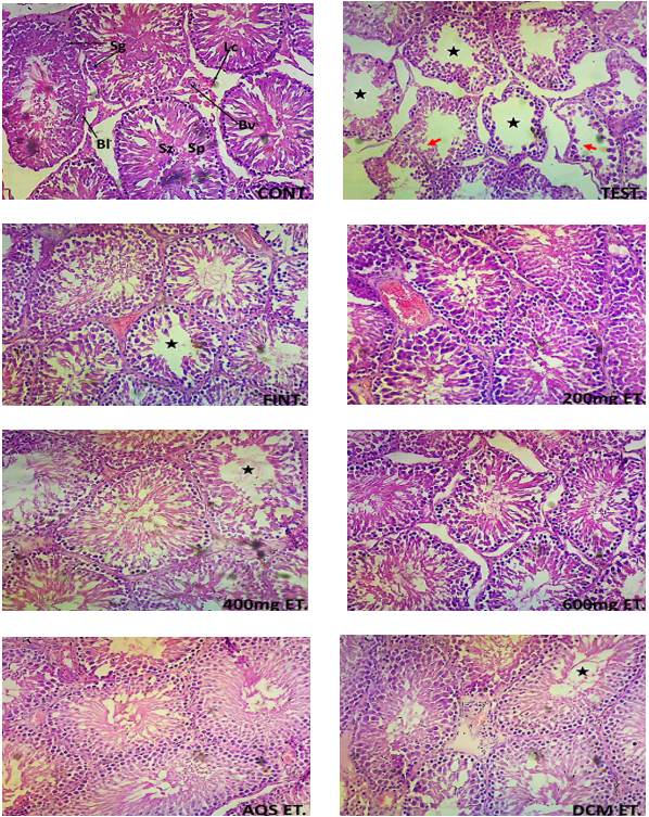
Histological sections of testes of rats receiving various treatments at magnification (x100) stained with H& E method revealed that rats in group 1 (normal control (CONT) treated with distilled water (10 mL/kg) had section demonstrating normal histo-architecture with well-presented seminiferous tubules having well-defined basement layer, well-lined spermatogenic cells and arrays of spermatozoa within the tubular lumen, and well-presented Leydig cells and blood vessels within the interstitial connective tissue. Group 2 (TEST) rats administered with testosterone only, 3 mg/kg, had sections revealing severe histo-architectural alteration, with seminiferous tubules with absence of spermatogenic lining cells, absence of spermatids and areas of degenerating spermatogenic cells, area of atrophied and scanty leydig cells, with widened tubular lumen having scanty presence of spermatozoa within the seminiferous tubules. Rats in group 3 (FINT) treated with finasteride, 5 mg/kg and testosterone, 3 mg/kg had testes sections demonstrating moderately histo-architectural alteration, with areas of altered spermatogenic processes, with widened tubular lumen within the seminiferous tubules. This was considered to be moderately affected. Rats in group 4 (200 mg ET) treated with extract, 200 mg/kg and testosterone, 3 mg/kg had sections demonstrating normal histo-architecture with well-presented seminiferous tubules having well-defined basement layer, well-lined spermatogenic cells and arrays of spermatozoa within the tubular lumen, and well-presented Leydig cells and blood vessels within the interstitial connective tissue. Group 5 (400 mg ET) treated with extract, 400 mg/kg and testosterone 3 mg/kg had sections demonstrating moderate histo-architectural alteration, with areas of altered spermatogenic processes, with widened tubular lumen within the seminiferous tubules. Testes sections of rats in group 6 (600 mg ET) treated with extract, 600 mg/kg and testosterone 3 mg/kg and group 7 (AQS ET) rats treated with aqueous fraction, 400 mg/kg and testosterone 3 mg/kg showed normal histo-architecture with well-presented seminiferous tubules having well-defined basement layer, well-lined spermatogenic cells and arrays of spermatozoa within the tubular lumen, and well-presented Leydig cells and blood vessels within the interstitial connective tissue, while group 8 (DCM ET) rats treated with dichloromethane fraction 400 mg/kg and testosterone 3 mg/kg had testes sections demonstrating moderate histo-architectural alteration, with areas of altered spermatogenic processes, with widened tubular lumen within the seminiferous tubules (Figure 4).
Discussion
The root of H. africana which is used traditionally in the treatment of prostate disorders was evaluated for effects on testosterone propionate-induced benign prostatic hyperplasia in male rats. The results of the study showed that root extract and fractions of H. africana significantly reduced the testosterone propionate elevated prostate weights, serum PSA and testosterone levels in male rats with the aqueous fraction exerting the major effect. This result was corroborated by histological findings in the prostate sections of the male rats.
The prostate weight is used as an important marker of BPH development (45). Increased prostate weight is an indication of prostate disorder as reported in earlier published study (46). Prostate gland enlargement is defined by the proliferation of the gland's biological components, including stromal and epithelial cells (47). Based on the considerable increased of the prostate weight in the testosterone- induced BPH rats, the results of this study validated the findings of previous research investigations that established an increase in prostate size as a critical predictor of BPH development (48). Agents used in the treatment of BPH such as finasteride or other agents are known to decrease prostate weight. In this study, the male rats with untreated BPH were observed to show increased prostate weight compare to the control group. However, the root extract/fractions treated animals were found to have reduced prostate weight compared to testosterone only treated BPH group. Also root extract/fractions treated animals were found to have reduced prostate index values. Increases in prostate index have been associated with the development of BPH (49). These decreases in prostate index further support the antiprostatic activity of the root extract. These results indicate that H. africana root extract mitigated the prostatic growth and enlargement induced by testosterone. This could have resulted from the reported genotoxic and cytotoxic activities of H. africana root extract which demonstrated the ability of the extract to inhibit cell division and proliferation as well as cause cell death (32), highly supporting its previously reported anticancer activity (25). In addition, these findings were further supported by histological examination of the prostate tissue. The animals with untreated BPH presented with atrophying histo-structure of the prostatic gland, with hyperplastic glandular epithelial cells and fibrolysis of the fibro-muscular tissue within the prostatic stroma, depicting a severe effect in the prostate, whereas the animals with BPH that had been treated with the root extract and fractions showed mild or no glandular hyperplasia especially in the group treated with the DCM fraction of the root extract. These results suggested that H. africana root extract/fractions possess the potential to inhibit the progression of BPH induced by testosterone.
A glycoprotein called prostate sensitive antigen (PSA), which is present in serum, is a semi-quantitative marker of prostatic hyperplasia and a predictor of BPH (50). Abnormal serum PSA level is an indicator of prostatic disorder. Reduced prostatic hyperplasia is linked to lower PSA levels, and this has a direct impact on 5α-reductase inhibition (51). Elevated serum PSA level as observed in this study due to testosterone propionate injection (3 mg/kg) indicated successful induction of benign prostatic hyperplasia. Treatment of male rats with finasteride, H. africana root extract and fractions significantly lowered the serum PSA level of the extract/fractions treated groups as compared to the testosterone only group. Finasteride is a selective inhibitor of type II 5α-reductase that catalyses dihydrotestosterone (DHT) formation from testosterone (52). The administration of root extract and fractions of H. africana significantly lowered the prostatic condition close to normal as observed by decreases in serum PSA and testosterone levels as well as pathological signs in prostate histology of the treated rats especially in the DCM fraction treated group. The observed reduction in serum PSA may suggest the extract/fractions ability to inhibit 5α-reductase activity which converts testosterone (53). Previous works on C. sieberiana, Z. portoricensis and Anona muricata extracts activities on testosterone-induced BPH had shown similar inhibitory potentials (46, 53, 54).
Blood levels of free testosterone are suggested to contribute significantly in the development of BPH and is known to encourage the growth of prostate cells (55). In TP-induced BPH rats, the extract/fractions significantly and non dose-dependently inhibited the increase in testosterone levels. This may suggest the H. africana root extract and fractions inhibitory potential on the production of DHT in serum and the prostate, leading to the excretion of free testosterone. H. africana root extract/fractions can be developed to treat BPH condition. The results of this investigation corroborate those of a study by Ibukun et al. (56) on Annona muricata (Soursop), which reduced testicular toxicity and prostatic impairment in male rats with BPH induced by testosterone propionate were reported. The significant lowering of serum testosterone levels in the root extract/fractions-treated rats with BPH may be linked to improved clearing of unbound testosterone in the bloodstream, preventing the conversion of active DHT by 5 α-reductase (57, 58). The results of this study revealed that testosterone caused a significant increase in MDA and decreases in SOD, GPx, GSH and catalase, which suggest oxidative stress condition. Development and progression of BPH have been reported to involve oxidative stress mechanisms (4). The root extract and fractions of H. africana have been reported to exert cellular antioxidant, anticancer and immunomodulatory (22) as well as in vitro and cellular antioxidant activities (18, 25). H. africana root extract/fractions administration in this study was found to significantly elevate the levels of SOD, GPx, GSH and catalase, while decreasing MDA level. This could have been possible through the antioxidant potentials of the constituents of the extract/fractions as reported earlier by Okokon et al. (15, 18), thereby attenuating oxidative stress condition induced by testosterone. Moreso, inflammation has been implicated in the pathogenesis of prostatitis induced by testosterone propionate (59). The observed antiprostatic activities of the root extract and fractions, as seen in the reduced levels of PSA and testosterone as well as pathological signs in the prostate tissue, could have resulted from the anti-inflammatory, cellular antioxidant and immunomodulatory activities of the root extract and fractions as earlier reported by Okokon et al., (12, 18, 22) and Umoh et al., (25). These actions may have contributed to the ameliorative/preventive activities of the root extract/fractions of H. africana against testosterone-induced BPH as evidenced in this study.
Dislipidemia has been associated with the pathogenesis and progression of BPH (60) and there is a scientific report on the correlation between hyperlipidemia and benign prostatic hyperplasia (61). Recent clinical and basic research evidence has demonstrated a possible linkage of cholesterol to benign prostatic hyperplasia. Accumulation of cholesterol within the lipid raft component of the cellular plasma membrane may stimulate signaling pathways that promote prostate tumor growth and progression (62). In this study, administration of testosterone was observed to caused significant elevation in levels of total cholesterol, triglyceride, low density lipoprotein and very low density lipoprotein, while the high-density lipoprotein level was not significantly reduced. These lipids parameters were reduced significantly (p < 0.05-0.001) when compared to testosterone only treated group following concomitant treatment with root extract and fractions of H. africana and finasteride. Although the mechanism in which dislipidaemia contributes to BPH progression has not been fully elucidated, studies have associated high triglyceride levels with prostate inflammation which can ultimately lead to BPH (60). Furthermore, Kayode et al. (63) reported alteration in the biochemical and lipid profile of testosterone propionate-induced benign prostatic hyperplasia in male Wistar rats treated with ketogenic diet. The elevated serum triglyceride concentration in the testosterone only treated BPH group relative to the normal control, positive control and extract/fractions-treated groups indicates the adverse effect of BPH on lipid metabolism, which promoted hyperlipidemia in the BPH rats. The elevated serum triglyceride level in the BPH untreated group agrees with Uroko et al. (64), who reported that high serum triglyceride level is associated with BPH pathogenesis. The extract’s reduction of the hyperlipidemia induced by testosterone further indicates and supports the antiprostatic potentials of the root extract/ fractions of H. africana against testosterone induced BPH as observed in this study which is largely due to the activities of the phytochemical constituents.
Evaluation of H. africana root extract/fractions on the semen of testosterone propionate-induced benign prostatic hyperplasia in male rats revealed that H. africana root has little or no toxic effect on testes and fecundity of adult male Wistar rats as demonstrated in the significant (p < 0.05) increased epididymal sperm count, sperm motility and live spermatozoa along with a simultaneous decrease in dead spermatozoa as compared to the rats of the group treated with testosterone only. This was further supported by the histological study of the testes whereby mild or no pathological effect was observed in the root extract/fractions treated rats compared to testosterone only treated group. This result corroborates earlier reported testicular protective potential of the root extract of H. africana (65).
Previous phytochemical analysis of dichloromethane fraction of H. africana by Okokon et al (18) showed that the root fraction contain pharmacologically active compounds such as dehydromevalonic lactone, phenol, 3, 4-dimethoxy-, phenol, 3, 4, 5-trimethoxy-, Xi-Germacr-9-en-12-oic acid, 6a - hydroxy-1-oxo-, c-lactone,(11S)-, dehydroxy-3-deoxy, 2 (1H) phenanthrenone, 3, 4, 4a, 9, 10, 10a hexahydro-6-methoxy-1, 1, 4a-trimethyl-7-(1-methylethyl)-, (4aS-trans)-, Retinoic acid, methyl ester, phenanthrene, 1, 2, 3, 4, 4a, 9, 10, 10a-octahydro-6-methoxy-1, 1, 4a-trimethyl7-(1-methyl ethyl)-,(4aS-trans)-, Myrcene, a-Phellandrene, 4a (2H) Phenanthrene carboxaldehyde, 1, 3, 4, 9, 10, 10a-hexahydro-6- methoxy-1, 1-dimethyl-7-(1-methyl ethyl) (4aS-trans)-, 4a (2H)-Phenanthrenemethanol, 1, 3, 4, 9, 10, 10a-hexahydro-6- methoxy-1, 1-dimethyl-7-(-1-methylethyl)-, (4aS-trans)-, limonene, terpinolene, linalool, a-terpineol among others. These compounds may have been responsible for the observed activities in this study.
Conclusion
The results of this study suggest strongly that the root extract and fractions of H. africana possess antiprostatic activity against testosterone-induced BPH through the antioxidant and anti-inflammatory activities of its phytochemical constituents.
Declarations
Conflict of Interest
The authors declare no conflicting interest.
Data Availability
The data generated during and/or analyzed during the current study are available from the corresponding author on reasonable request.
Ethics Statement
All animal experiments were approved by the Institutional Animal Care and Use Committee / Animal Research Ethics Committee of University of UYO with approval no. (UU/CHS/IHREC/25/VOL.1/14) and conducted in accordance with relevant guidelines and regulations.
Funding Information
The author(s) declare that no financial support was received for the research, authorship, and/or publication of this article.
References
- Foo KT. Pathophysiology of clinical benign prostatic hyperplasia. Asian Journal of Urology. 2017;4(3):152-157. doi: https://doi.org/10.1016/j.ajur.2017.06.003
- Lim KB. Epidemiology of clinical benign prostatic hyperplasia. Asian Journal of Urology. 2017;4(3):148-151. doi: https://doi.org/10.1016/j.ajur.2017.06.004
- Madersbacher S, Sampson N, Culig Z. Pathophysiology of Benign Prostatic Hyperplasia and Benign Prostatic Enlargement: A Mini-Review. Gerontology. 2019;65(5):458-464. doi: https://doi.org/10.1159/000496289
- Baral N, Paudel B, Das B, Aryal M, Gautam A, Lamsal M. Preparing tutors for problem-based learning: An experience from B. P. Koirala Institute of Health Sciences, Nepal. Kathmandu Univ. Med. J. 1970;8(1):141-145. doi: https://doi.org/10.3126/kumj.v8i1.3237
- Chandra K, Salman AS, Mohd A, Sweety R, Ali KN. Protection against FCA induced oxidative stress induced DNA damage as a model of arthritis and In vitro anti-arthritic potential of costusspeciosus rhizome extract. International Journal of Pharmacognosy and Phytochemical Research, 2015;7(2): 383-389.
- Zhang M, Luo C, Cui K, Xiong T, Chen Z. Chronic inflammation promotes proliferation in the prostatic stroma in rats with experimental autoimmune prostatitis: study for a novel method of inducing benign prostatic hyperplasia in a rat model. World J Urol. 2020;38(11):2933-2943. doi: https://doi.org/10.1007/s00345-020-03090-6
- Basalingappa KM, Anitha B, Raghu N, Gopenath TS, Karthikeyan M, Gnanasekaran A, et al. Medicinal Uses of Carica Papaya. Jonam. 2018;2(6):1-11. doi: https://doi.org/10.23880/jonam-16000144
- Hutchinson J, Dalziel JM. Flora of West Tropical Africa. 2nd edition. Crown Agents for Overseas Government and Administration, Vol.1, Part 2, p.638,1973.
- Okokon JE, Ita BN, Udokpoh AE. The in-vivo antimalarial activities ofUvaria chamaeandHippocratea africana. Annals of Tropical Medicine & Parasitology. 2006;100(7):585-590. doi: https://doi.org/10.1179/136485906x118512
- Etukudo I. Forests: Our Divine Treasure. Dorand Publishers, Nigeria, p. 156 – 180,2000.
- Etukudo I. Ethnobotany: Conventional and Traditional Uses of Plants.The Verdict Press, Nigeria, p. 83 – 134,2003.
- Ajibesin KK, Ekpo BA, Bala DN, Essien EE, Adesanya SA. Ethnobotanical survey of Akwa Ibom State of Nigeria. Journal of Ethnopharmacology. 2008;115(3):387-408. doi: https://doi.org/10.1016/j.jep.2007.10.021
- Okokon JE, Amechi P, Udobang JA, Bankehde HK, Bassey AL. Cytotoxic, in vitro and in vivo antimalarial activities of Hippocratea africana.Souith Asian J Parasitol 2021;5(4):1-14.
- Okokon JE, Antia BS, Umoh E. Analgesic and Anti-Inflammatory Effects of Ethanolic Root Extract of Hippocratea africana. International J. of Pharmacology. 2007;4(1):51-55. doi: https://doi.org/10.3923/ijp.2008.51.55
- Okokon JE, Antia BS, Umoh EE, Etim EI. Antidiabetic and hypolipidaemic activities of Hippocratea africana. Int J Drug Dev Res, 2010; 2: 501 -506.
- Okokon JE, Chinyere PC, Amaechi P, Bassey AL, Thomas PS. Antioxidant, antidiabetic and hypolipidemic activities of ethanol root extract and fractions of Hippocratea africana. Trop J Nat Prod Res. 2022;6(3):446-453.
- Okokon JE, Akpan HD, Ekaidem I, Umoh EE. Antiulcer and antidiarrheal activity of Hippocratea africana. Pak J Pharm Sci 2011;24:201- 205.
- Okokon JE, Nwafor PA, Charles U, Dar A, Iqbal Choudhary M. Antioxidative burst and hepatoprotective effects of ethanol root extract ofHippocratea africanaagainst paracetamol-induced liver injury. Pharmaceutical Biology. 2013;51(7):872-880. doi: https://doi.org/10.3109/13880209.2013.768273
- Okokon JE, Anagboso MO, Noah KU. Hippocratea africana Root Extract and Fractions Attenuated Carbon Tetrachloride-Induced Oxidative Stress and Liver Injuries in Rats. J. Compl. Altern. Med. Res. 2024;25(8):71-83. doi: https://doi.org/10.9734/jocamr/2024/v25i8561
- Noah K, Anagboso MO, Iyanyi UL, Ajaghaku DL, Okokon JE. Hippocratea africana Root Extract and Fractions Ameliorates Paracetamol (PCM)-Induced Oxidative Stress and Liver Injuries in Rats. Ajbgmb. 2023;15(3):114-122. doi: https://doi.org/10.9734/ajbgmb/2023/v15i3344
- Joseph O, Okokon J, Okokon J. Hepatoprotective activity of ethanol root extract and fractions of Hippocratea africana against doxorubicin-induced liver toxicity. jcbr. 2023;3(4, July-August):1132-1153. doi: https://doi.org/10.54117/jcbr.v3i4.6
- Okokon J, Choudhary M, Dar A. Immunostimulatory, cytotoxic and antileishmanial activity of Mammea africana from Nigeria. J Nat Pharm. 2012;3(2):105. doi: https://doi.org/10.4103/2229-5119.102754
- Okokon JE, Davies K, Okokon PJ, Antia BS. Depressant, anticonvulsant and antibacterial activities of Hippocratea africana. Int J Phytother 2014;4 (3):144 – 153.
- Okokon JE, Chinyere CP, Bassey AL, Udobang JA. In vivo alpha amylase and alpha glucosidase activities of ethanol root extract and fractions of Hippocratea africana. South Asian J Parasitol, 2021;5(4): 42-48.
- Umoh UF, Thomas PS, Essien EE, Okokon JE, De Leo M, Ajibesin KK, et al. Isolation and characterization of bioactive xanthones from Hippocratea africana (Willd.)Loes.ex Engl. (Celastraceae). Journal of Ethnopharmacology. 2021;280:114031. doi: https://doi.org/10.1016/j.jep.2021.114031
- Noah KU, Udobang JA, Okokon JE, Anagboso MO, Ebong NO. Nephroprotective Activities of Ethanol Root Extract and Fractions of Hippocratea africana Against Doxorubicin-Induced Kidney Toxicity. Biomednatproch. 2023;12(2):477-484. doi: https://doi.org/10.14421/biomedich.2023.122.477-484
- Noah K, Edem UA, Iyanyi UL, Ajaghaku DL, Okokon JE. Hippocratea africana root extract and fractions ameliorated carbon tetrachloride-induced oxidative stress and kidney injuries in rats. Asian J Nat Prod Biochem. 2024;22(2). doi: https://doi.org/10.13057/biofar/f220201
- Noah K, Edem UA, Iyanyi UL, Ajaghaku DL, Okokon JE. Hippocratea africana root extract and fractions ameliorated carbon tetrachloride-induced oxidative stress and kidney injuries in rats. Asian J Nat Prod Biochem. 2024;22(2). doi: https://doi.org/10.13057/biofar/f220201
- Johnny, I. I., Okokon, J. E., Ochigbo, E. B., Udo, I. J., Adefabi, A. M. Genotoxic and Cytotoxic Activities of Hippocratea africana Loes. Ex. Engl. Celastraceae Root Extract. Ajbgmb. 2023;15(2):38-45. doi: https://doi.org/10.9734/ajbgmb/2023/v15i2331
- Noah KU, Okokon JE. Hippocratea africana Ethanol Root Extract and Fractions Attenuate Doxorubicin-Induced Testicular Toxicity and Oxidative Stress. Biomednatproch. 2024;13(2):397-406. doi: https://doi.org/10.14421/biomedich.2024.132.397-406
- Asanga EE, Noah KU, Okokon JE, Ekeleme CM, Udoh IE, Bassey A, et al. Integrative investigation on Hippocratea africana root: insights from cardio-protective, anti-oxidative stress activities, isolation, GC/MS, and pharmacological significance profiling. BMC Complement Med Ther. 2025;25(1). doi: https://doi.org/10.1186/s12906-025-04941-8
- Okokon JE, Okokon PJ, Sahal D. In vitro antiplasmodial activity of some medicinal plants from Nigeria. Int J Herbal Med 2017;5 (5):102-109.
- Sasidharan S, Kp S, Bhaumik A, Kanti Das S, Nair J H. Administration of Caesalpinia bonduc Seed Extracts Ameliorates Testosterone-Induced Benign Prostatic Hyperplasia (BPH) in Male Wistar Rats. Rru. 2022;Volume 14:225-239. doi: https://doi.org/10.2147/rru.s365598
- Kenjale R, Shah R, Sathaye S. Effects of Chlorophytum borivilianum on sexual behaviour and sperm count in male rats. Phytotherapy Research. 2008;22(6):796-801. doi: https://doi.org/10.1002/ptr.2369
- Yokoi K, Uthus EO, Nielsen FH. Nickel Deficiency Diminishes Sperm Quantity and Movement in Rats. Bter. 2003;93(1-3):141-154. doi: https://doi.org/10.1385/bter:93:1-3:141
- Filler R. Methods for Evaluation of Rat Epididymal Sperm Morphology. Elsevier; 1993. doi: https://doi.org/10.1016/b978-0-12-461207-5.50025-0
- Tietz NW. Fundamentals of Clinical Chemistry, 2nd ed. W.B. Saunders Co, Philadelphia, P.A. p.335- 1208,1976.
- Friedewald WT, Levy RI, Fredrickson DS. Estimation of the Concentration of Low-Density Lipoprotein Cholesterol in Plasma, Without Use of the Preparative Ultracentrifuge. Clinical Chemistry. 1972;18(6):499-502. doi: https://doi.org/10.1093/clinchem/18.6.499
- Esterbauer H, Cheeseman KH. [42] Determination of aldehydic lipid peroxidation products: Malonaldehyde and 4-hydroxynonenal. Elsevier; 1990. doi: https://doi.org/10.1016/0076-6879(90)86134-h
- Marklund S, Marklund G. Involvement of the Superoxide Anion Radical in the Autoxidation of Pyrogallol and a Convenient Assay for Superoxide Dismutase. European Journal of Biochemistry. 1974;47(3):469-474. doi: https://doi.org/10.1111/j.1432-1033.1974.tb03714.x
- Sinha AK. Colorimetric assay of catalase. Analytical Biochemistry. 1972;47(2):389-394. doi: https://doi.org/10.1016/0003-2697(72)90132-7
- Lawrence RA, Burk RF. Glutathione peroxidase activity in selenium-deficient rat liver. Biochemical and Biophysical Research Communications. 1976;71(4):952-958. doi: https://doi.org/10.1016/0006-291x(76)90747-6
- Ellman GL. Tissue sulfhydryl groups. Archives of Biochemistry and Biophysics. 1959;82(1):70-77. doi: https://doi.org/10.1016/0003-9861(59)90090-6
- Drury RA, Wallington EA. Carleton's Histological Techniques. 5th Edition, Oxford University Press, New York, p.195,1990.
- Lee MY, Shin IS, Seo CS, Lee NH, Ha HK, Son JK, et al. Effects of Melandrium firmum methanolic extract on testosterone-induced benign prostatic hyperplasia in Wistar rats. Asian J Androl. 2012;14(2):320-324. doi: https://doi.org/10.1038/aja.2011.166
- Leje I, U. W, Yeldu M, Alhassan H, Wasagu IZ, Abubakar U, et al. Effects of Aqueous Fruit Extract of Annona muricata on Testosterone Propionate Induced Benign Prostate Hyperplasia (BPH) in Male Wistar Rats. Asian J. Res. Biochem. 2024;14(4):72-83. doi: https://doi.org/10.9734/ajrb/2024/v14i4295
- Kapoor R, Liatsikos EN, Badlani G. Endoprostatic stents for management of benign prostatic hyperplasia. Current Opinion in Urology. 2000;10(1):19-22. doi: https://doi.org/10.1097/00042307-200001000-00005
- Shin IS, Lee MY, Jung DY, Seo CS, Ha HK, Shin HK. Ursolic acid reduces prostate size and dihydrotestosterone level in a rat model of benign prostatic hyperplasia. Food and Chemical Toxicology. 2012;50(3-4):884-888. doi: https://doi.org/10.1016/j.fct.2012.01.007
- Barry MJ, Fowler FJ, O’Leary MP, Bruskewitz RC, Holtgrewe HL, Mebust WK, et al. The American Urological Association Symptom Index for Benign Prostatic Hyperplasia. Journal of Urology. 1992;148(5 Part 1):1549-1557. doi: https://doi.org/10.1016/s0022-5347(17)36966-5
- Morcos MA, Afifi NM. Effect of doxazocin on experimentally induced prostatic hyperplasia in adult male albino rats. The Egyptian Journal of Histology. 2011;34(4):870-882. doi: https://doi.org/10.1097/01.ehx.0000407624.42021.3b
- Sing B, Ram SN, Pandey VB, Joshi VK, Gambhir SS. Studies on antiinflammatory activity of taraxasterol acetate from Echinops echinatus in rats and mice. Phytotherapy Research. 1991;5(3):103-106. doi: https://doi.org/10.1002/ptr.2650050303
- Traish AM, Morgentaler A. RE: Effect of finasteride on serum levels of androstenedione, testosterone and their 5α-reduced metabolites in men at risk for prostate cancer. The Journal of Steroid Biochemistry and Molecular Biology. 2013;138:462. doi: https://doi.org/10.1016/j.jsbmb.2013.06.013
- Ekeyi Y, Uchendu NO, Anaduaka EG, Ezeanyika LUS. Ethanol extract of Cassia sieberiana leaves ameliorates deviances associated with benign prostatic hyperplasia in rats. All Life. 2021;14(1):473-483. doi: https://doi.org/10.1080/26895293.2021.1927857
- Kayode, Dasofunjo. Effect of ethanol extract of Zapoteca portoricensis stem on liver, kidney and prostatic biomarkers of testosterone-induced benign prostate hyperplasia (BPH) in male albino rats. Aust. J. Basic & Appl. Sci. 2018. doi: https://doi.org/10.22587/ajbas.2018.12.10.4
- Mulia K, Winarcahyo SW, Krisanti E, Kurniasuci D. Practical Isolation of Bullatacin from <i>Annona muricata</i> Leaves Extract Using an Open Column Chromatography Technique. Amr. 2013;789:545-550. doi: https://doi.org/10.4028/www.scientific.net/amr.789.545
- Ibukun OE, Ogunlade LO, Oladipo GO. Annona muricata (Soursop) mitigated testicular toxicity and prostatic impairment in testosterone-propionate-induced BPH in Male Rats. Achievers J Scientific Res. 2023;5(2):01-11.
- Liu J, Fang T, Li M, Song Y, Li J, Xue Z, et al. Pao Pereira Extract Attenuates Testosterone-Induced Benign Prostatic Hyperplasia in Rats by inhibiting 5α-Reductase. Sci Rep. 2019;9(1). doi: https://doi.org/10.1038/s41598-019-56145-z
- Roehrborn CG, Nuckolls JG, Wei JT, Steers W. The Benign Prostatic Hyperplasia Registry and Patient Survey: study design, methods and patient baseline characteristics. BJU International. 2007;100(4):813-819. doi: https://doi.org/10.1111/j.1464-410x.2007.07061.x
- Krušlin B, Tomas D, Džombeta T, Milković-Periša M, Ulamec M. Inflammation in Prostatic Hyperplasia and Carcinoma—Basic Scientific Approach. Front. Oncol. 2017;7. doi: https://doi.org/10.3389/fonc.2017.00077
- Gacci M, Corona G, Sebastianelli A, Serni S, De Nunzio C, Maggi M, et al. Male Lower Urinary Tract Symptoms and Cardiovascular Events: A Systematic Review and Meta-analysis. European Urology. 2016;70(5):788-796. doi: https://doi.org/10.1016/j.eururo.2016.07.007
- Erbay G, Ceyhun G. Association between hyperlipidemia and prostatic enlargement: A case-control study. Urologia. 2021;89(1):58-63. doi: https://doi.org/10.1177/03915603211003401
- Tong YC. The role of cholesterol in prostatic diseases. Urological Science. 2011;22(3):97-102. doi: https://doi.org/10.1016/j.urols.2011.08.002
- Kayode OT, Owolabi AV, Kayode AAA. Biochemical and histomorphological changes in testosterone propionate-induced benign prostatic hyperplasia in male Wistar rats treated with ketogenic diet. Biomedicine & Pharmacotherapy. 2020;13:110863. doi: https://doi.org/10.1016/j.biopha.2020.110863
- Uroko RI, Uche ME, Nweje-Anyalowu PC, Obiwuru I, Aguwamba C, Aaron CF. Combined Anthocleista vogelii and Alstonia boonei Stem Barks Extract Alleviates Hyperlipidaemia and Renal Malfunctions in Benign Prostatic Hyperplasia-Induced Rats. Biomednatproch. 2022;12(1):69-78. doi: https://doi.org/10.14421/biomedich.2023.121.69-78
- Noah KU, Okokon JE. Hippocratea africana Ethanol Root Extract and Fractions Attenuate Doxorubicin-Induced Testicular Toxicity and Oxidative Stress. Biomednatproch. 2024;13(2):397-406. doi: https://doi.org/10.14421/biomedich.2024.132.397-406