RESEARCH ARTICLE
Breadfruit Peel Derived Pectin as a Natural Polymer for Gastroretentive Floating Beads of Amoxicillin: Formulation and Characterization
Academic Editor: Rifka Nurul Utami
Sciences of Pharmacy|Vol. 5, Issue 3, pp. 332-342 (2026)
CC BY 4.0-2026 Authors
Views
Downloads
Shares

Received
Apr 7, 2026Revised
Jun 22, 2026Accepted
Jul 8, 2026Published
Jul 20, 2026
Abstract
Amoxicillin is widely used to treat gastric infections; however, conventional oral dosage forms often exhibit rapid gastric emptying and short gastric residence time, which may reduce therapeutic effectiveness. Gastroretentive floating drug delivery systems can prolong gastric retention and provide sustained drug release. This study aimed to develop gastroretentive floating beads of amoxicillin using pectin extracted from breadfruit peel (Artocarpus altilis) and compare their characteristics with those prepared using commercial pectin. Floating beads were prepared by ionotropic gelation using calcium chloride as a crosslinking agent and sodium bicarbonate as a gas-forming agent. The beads were characterized for morphology, floating behavior, entrapment efficiency, and drug release. All evaluations were performed in triplicate (n = 3). The prepared beads exhibited spherical morphology with diameters of 0.24–0.42 mm and weights of 9.26–11.38 mg. Floating lag time ranged from 10–22 seconds, and all formulations remained buoyant for up to 8 hours. Entrapment efficiency ranged from 31–47%, with formulations prepared using commercial pectin showing values of 34–47% and breadfruit peel-derived pectin showing values of 31–43%, with formulation F3 containing 3.5% commercial pectin showing the highest value. In vitro drug release studies demonstrated sustained release of amoxicillin for up to 8 hours, whereas conventional tablets released 64.77% of the drug within 2 hours. Drug release followed the Higuchi model (R² = 0.989–0.996), indicating diffusion-controlled release. These findings demonstrate that breadfruit peel-derived pectin is a promising natural polymer for gastroretentive floating bead formulations and exhibits performance comparable to commercial pectin.
Introduction
Amoxicillin is a widely prescribed β-lactam antibiotic for the treatment of various bacterial infections, including those associated with Helicobacter pylori. The therapeutic success of amoxicillin in H. pylori eradication depends largely on maintaining sufficient drug concentrations within the stomach, where the microorganism resides in the gastric mucus layer (1). However, conventional oral dosage forms are often limited by rapid gastric emptying and a relatively short residence time in the gastric environment, which may reduce local drug availability and therapeutic efficacy (2). To overcome these limitations, gastroretentive drug delivery systems (GRDDS) have been developed to prolong gastric residence and sustain drug release. Among the various GRDDS approaches, floating drug delivery systems have attracted considerable attention because they remain buoyant in gastric fluid, thereby enhancing gastric retention and improving the duration of drug exposure at the target site (3).
Natural polymers are increasingly utilized in controlled-release and gastroretentive formulations due to their biocompatibility, biodegradability, safety, and environmental sustainability. Pectin is one of the most promising natural polymers because of its ability to form gel matrices and crosslinked networks in the presence of divalent cations such as calcium. These properties make pectin particularly suitable for the development of floating and sustained-release drug delivery systems (4). Although commercial pectin has been extensively employed in pharmaceutical formulations, recent studies have explored alternative pectin sources obtained from agricultural by-products and fruit-processing waste. The utilization of such materials not only supports waste valorization and environmental sustainability but also offers a potentially economical source of pharmaceutical excipients.
Breadfruit peel (Artocarpus altilis), an abundant agricultural waste generated during fruit processing and consumption, has been reported as a potential source of pectin with favorable physicochemical characteristics. Previous investigations have primarily focused on the extraction process and characterization of breadfruit peel-derived pectin, while information regarding its application in gastroretentive floating drug delivery systems remains limited (5, 6). Furthermore, comparative studies evaluating the performance of breadfruit peel-derived pectin and commercial pharmaceutical-grade pectin under identical formulation conditions are scarce. Therefore, the present study aimed to formulate gastroretentive floating beads containing amoxicillin using pectin isolated from breadfruit peel and to compare their characteristics with those prepared using commercial pectin. The resulting formulations were evaluated in terms of physicochemical properties, buoyancy behavior, swelling capacity, drug entrapment efficiency, and in vitro drug release performance to determine the suitability of breadfruit peel-derived pectin as a sustainable alternative polymer for gastroretentive drug delivery applications.
Methodology
Study Design
This experimental laboratory study aimed to isolate pectin from breadfruit peel and evaluate its suitability as a matrix-forming polymer in the preparation of gastroretentive floating beads containing amoxicillin. The extracted pectin was characterized using FT-IR spectroscopy and compared with commercial pectin. Floating beads were formulated using both polymer sources and evaluated for physicochemical properties, buoyancy behavior, swelling characteristics, drug entrapment efficiency, release kinetics, and in vitro drug release profile. All formulations and evaluations were performed in triplicate (n = 3) to ensure experimental reproducibility.
Materials
Fresh breadfruit peel was obtained from local cultivation areas in Serdang Bedagai, North Sumatra, Indonesia. Amoxicillin trihydrate was used as the model drug (Sigma-Aldrich, USA). Commercial pharmaceutical-grade pectin (Sigma-Aldrich, USA) was used as the reference polymer. Calcium chloride (Merck, Germany) was employed as a crosslinking agent. Other reagents included citric acid (Merck, Germany), sodium bicarbonate (Merck, Germany), sodium hydroxide (Merck, Germany), phenolphthalein indicator (Merck, Germany), hydrochloric acid (Merck, Germany), and phosphate buffer components including potassium dihydrogen phosphate and disodium hydrogen phosphate (Merck, Germany). Ethanol 96% (Brataco Chemical, Indonesia) and other analytical-grade solvents were also used. All chemicals were of analytical grade and used without further purification. Distilled water (Aqua Destillata, Ikapharmindo, Indonesia) was used throughout the study.
Plant Identification
Breadfruit samples were taxonomically identified at Universitas Sumatera Utara to confirm plant authenticity, with identification number 050/MEDA/2025.
Preparation of Simplicia
The peel was separated, washed thoroughly to remove adhering impurities, and cut into small pieces. Drying was conducted under controlled conditions until constant weight was achieved. The dried material was then pulverized into fine powder and stored in airtight containers for further use (7).
Isolation of Pectin
The pectin isolation procedure was carried out following the method described by (8), with minor modifications. A total of 90 g of powdered breadfruit peel was extracted using 1000 mL of 7.5% citric acid solution in a beaker glass. The mixture was continuously stirred and heated at 70–80 °C for 90 min under constant stirring conditions to facilitate the extraction process. After extraction, the mixture was allowed to cool and then filtered through a muslin cloth to obtain the pectin filtrate. Pectin was precipitated by adding 96% ethanol at a ratio of 1:1.5 (v/v). The resulting pectin coagulum was separated by filtration using filter paper. The wet pectin was washed with 96% ethanol to remove residual acid. The purified wet pectin was then dried in an oven at 50 °C for 12 h. The dried pectin was subsequently ground and sieved to obtain a uniform powder. To obtain sufficient yield, the extraction process was repeated as necessary.
Preparation of Amoxicillin Gastroretentive Floating Beads
Amoxicillin gastroretentive sustained-release floating beads were prepared using both isolated pectin from breadfruit peel and commercial pectin at different concentrations, as shown in Table 1 (8). The selected pectin concentrations (3.0%, 3.25%, and 3.5% w/v) were determined based on preliminary optimization studies conducted prior to the main experiment. Preliminary trials indicated that pectin concentrations below 3.0% produced beads with inadequate structural integrity and poor shape retention, whereas concentrations above 3.5% generated highly viscous dispersions that were difficult to process and resulted in non-uniform bead formation. Therefore, the concentration range of 3.0–3.5% was considered appropriate for further formulation and evaluation. Polymer solutions were prepared by dissolving pectin at concentrations of 3.0%, 3.25%, and 3.50% (w/v) in 10 mL of distilled water. Sodium bicarbonate (NaHCO₃), serving as a gas-forming agent, was added at a fixed amount of 50 mg and stirred until uniformly dispersed. Amoxicillin (500 mg) was then incorporated into the polymer mixture, followed by sonication for 30 min to ensure homogeneous dispersion. The resulting dispersion was carefully dropped into 20 mL of a crosslinking medium consisting of 2% (w/v) calcium chloride prepared in 10% (v/v) acetic acid under continuous stirring. Ionic gelation occurred immediately upon contact with the crosslinking medium, resulting in the formation of floating beads. The acidic medium was incorporated to facilitate the reaction between acetic acid and sodium bicarbonate, thereby generating carbon dioxide gas responsible for bead buoyancy. Ionic gelation occurred immediately upon contact with the crosslinking solution, resulting in the formation of floating beads. The beads were allowed to remain in the calcium chloride solution for 10–15 min to ensure adequate curing and stabilization of the polymeric network.
Subsequently, the beads were collected by filtration using filter paper and gently blotted to remove excess solution. The beads were rinsed with distilled water to remove residual reagents, blotted again using filter paper, and dried in an oven at 50–60 °C for 60 min. The dried beads were immediately subjected to characterization and evaluation. The composition of each formulation used in this study is presented in Table 1.
| Materials | Formulation | |||||
|---|---|---|---|---|---|---|
| F1 | F2 | F3 | F4 | F5 | F6 | |
| Amoxicillin (mg) | 500 | 500 | 500 | 500 | 500 | 500 |
| Commercial pectin (%) | 3.0 | 3.25 | 3.50 | - | - | - |
| Isolated pectin (%) | - | - | - | 3.0 | 3.25 | 3.50 |
| NaHCO3 (mg) | 50 | 50 | 50 | 50 | 50 | 50 |
| CaCl2 (%) | 2 | 2 | 2 | 2 | 2 | 2 |
| Note: All formulations were gelled in 20 mL of 2% (w/v) CaCl₂ containing 10% (v/v) acetic acid. | ||||||
Evaluation of Floating Beads
Determination of Bead Diameter
Bead diameter was determined using a digital caliper. Five randomly selected beads were measured from each replicate batch. This sample size was selected for preliminary physicochemical characterization and was consistently applied across all formulations, and the experiment was performed in triplicate (n = 3). The results are expressed as mean ± standard deviation (SD) (9).
Determination of Bead Weight
Bead weight was determined using an analytical balance. Five randomly selected beads were weighed from each replicate batch, and the experiment was performed in triplicate (n = 3). The results are expressed as mean ± standard deviation (SD) (9).
Floating Lag Time and Floating Duration Study
Floating behavior of the beads was evaluated by determining both the floating lag time and floating duration. A total of ten beads were placed in a beaker containing simulated gastric fluid (pH 1.2). The floating lag time was recorded as the time required for the beads to rise to the surface of the medium. The floating duration was defined as the total time during which the beads remained buoyant and was observed at hourly intervals for up to 8 h. This test was conducted to assess the buoyancy characteristics and gastroretentive potential of the floating bead formulations (10). The experiment was performed in triplicate (n = 3), and the reported floating lag time values represent the mean values obtained from three independent measurements.
Swelling Study of Amoxicillin Floating Beads
The swelling behavior of amoxicillin floating beads was evaluated to determine the expansion properties of the beads in simulated gastric conditions. A total of 100 mg of dried floating beads was accurately weighed (W₁) and placed into 900 mL of simulated gastric fluid (pH 1.2) maintained at 37 ± 0.5 °C using a dissolution apparatus operated at 50 rpm. At predetermined time intervals, the beads were removed, filtered using filter paper, and weighed again (W₂) after removing excess surface liquid. The swelling index was calculated based on the increase in bead weight after immersion in the medium. This test was conducted for 5 h to evaluate the swelling characteristics of the floating beads (11). All measurements were performed in triplicate (n = 3), and the results were expressed as mean ± standard deviation. The swelling index was calculated using the Equation 1, where W1 = Initial weight of beads and W2 = Weight of beads after swelling.
Determination of Amoxicillin Entrapment Efficiency in Floating Beads
A total of 20 mL of calcium chloride filtrate obtained from the filtration of the amoxicillin floating bead preparation was transferred into a 50 mL volumetric flask and diluted to volume with simulated gastric fluid (pH 1.2). The solution was then filtered, and 1 mL of the filtrate was further transferred into a 25 mL volumetric flask and diluted to the mark with simulated gastric fluid (pH 1.2). The concentration of unentrapped amoxicillin in the filtrate was determined using an ultraviolet spectrophotometer at a maximum wavelength of 228 nm. The amount of drug entrapped in the floating beads was calculated by subtracting the amount of free drug from the initial drug content (8). The entrapment efficiency determination was performed in triplicate (n = 3), and the results were expressed as mean ± standard deviation. The % of drug entrapment efficiency was calculated using the Equation 2, where W2 = total drug amount and W1 = free drug amount in the supernatant.
Amoxicillin Release Study from Floating Bead Formulations
In Vitro Release Study of Amoxicillin
The in vitro release of amoxicillin from the floating bead formulation was evaluated using a paddle-type dissolution apparatus. The test was conducted in 900 mL of simulated gastric fluid (pH 1.2) maintained at 37 ± 0.5 °C, with a stirring speed of 50 rpm. Floating beads equivalent to 500 mg of amoxicillin were placed into the dissolution vessel after the temperature and agitation speed had stabilized. Aliquots of 5 mL were withdrawn at predetermined time intervals of 5, 10, 15, 20, 30, 45, 60, 90, 120, 150, 180, 240, 300, 360, 420, and 480 min. Each withdrawn sample was diluted with simulated gastric fluid (pH 1.2) to a final volume of 100 mL prior to analysis. To maintain a constant dissolution volume, each withdrawn sample was immediately replaced with an equal volume (5 mL) of fresh simulated gastric fluid at the same temperature. The concentration of released amoxicillin was determined using ultraviolet spectrophotometry at a wavelength of 228 nm. Each formulation was tested in triplicate to ensure reproducibility of the results (12).
Analysis of Amoxicillin Release Kinetics
The dissolution profiles obtained from each formulation were analyzed by fitting the release data to several mathematical kinetic models to determine the mechanism of drug release. The models applied included zero-order kinetics, first-order kinetics, Higuchi model, and Korsmeyer–Peppas model. The zero-order model describes drug release at a constant rate independent of drug concentration, while the first-order model explains concentration-dependent drug release. The Higuchi model characterizes drug release from matrix systems based on diffusion mechanisms, whereas the Korsmeyer–Peppas model is used to identify the release mechanism when more than one type of release phenomenon is involved. The most appropriate release model was determined based on the highest correlation coefficient (R²) value. This analysis was performed to identify the release kinetics and to elucidate the mechanism governing amoxicillin release from the gastroretentive floating bead system (13).
Statistical Analysis
All experiments were performed in triplicate (n = 3) and results were expressed as mean ± standard deviation (SD). Statistical comparisons of cumulative drug release among formulations were analyzed using one-way analysis of variance (ANOVA) followed by Tukey's post hoc multiple comparison test. Statistical significance was established at p < 0.05. Confidence intervals (95% CI) were calculated where applicable.
Results and Discussion
Sample Preparation Result
Fresh breadfruit was collected from Martebing, Serdang Bedagai, with an initial weight of 1 kg. The breadfruit peel was separated, washed, dried, and subsequently ground into powder to obtain simplicia. After the drying and milling process, 90 g of powdered breadfruit peel simplicia were obtained. The loss on drying of the breadfruit peel was found to be 8%, while the simplicia yield was calculated to be 8.32%. The powdered simplicia was then subjected to pectin isolation using 7.5% citric acid as the extraction solvent. From 90 g of breadfruit peel powder, 7.41 g of pectin was obtained, corresponding to a pectin yield of approximately 8%. The obtained yield indicates that breadfruit peel has potential as an alternative natural source of pectin. The extraction yield is influenced by several factors, including extraction temperature, extraction time, pH, and solvent concentration. Acidic extraction using citric acid facilitates the hydrolysis of protopectin into soluble pectin, thereby improving extraction efficiency. In addition, the moderate extraction temperature helps preserve pectin structure and prevents excessive degradation during the isolation process. These results suggest that breadfruit peel can be effectively utilized as a natural polymer source for pharmaceutical applications, particularly in gastroretentive floating bead formulations.
Evaluation Results and Characteristics of the Formulation
Determination of Bead Specifications
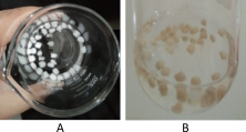
The beads were produced by dropping a mixture of pectin solution and drug into a calcium chloride solution. The interaction between pectin and calcium ions forms an “egg-box” structure, which enables the entrapment of the drug within the polymeric matrix (8). The specification assessment of pectin beads containing amoxicillin included observations of shape, color, as well as measurements of bead weight and diameter. Visual evaluation of all six formulations showed that the beads were spherical or nearly spherical in shape. Beads prepared using commercial pectin (F1–F3) appeared white, whereas beads formulated with pectin isolated from breadfruit peel exhibited a yellowish-brown color. This difference in color is attributed to the inherent color variation between pectin derived from breadfruit peel and commercial pectin. Isolated breadfruit peel pectin typically appears yellowish-brown, while commercial pectin is generally white to pale yellow (5). The morphological appearance of beads from each formulation is presented in Figure 1.
Bead Diameter and Weight Results
The diameter of beads prepared using commercial pectin ranged from 0.24 to 0.40 mm, whereas beads formulated with breadfruit peel-derived pectin ranged from 0.26 to 0.42 mm. Overall, no substantial difference was observed in bead size between formulations prepared with commercial pectin and those prepared with isolated breadfruit peel pectin. However, pectin concentration influenced bead diameter, with higher concentrations associated with larger bead diameters in both commercial and breadfruit peel pectin formulations. This trend may be related to differences in the rheological properties of the polymer dispersion during bead formation, although viscosity was not directly measured in the present study (11). The bead diameter for each formulation is presented in Table 2.
| Formulation | Average Diameter (mm) ± SD |
|---|---|
| F1 (Commercial Pectin 3%) | 0.24 ± 0.05 |
| F2 (Commercial Pectin 3.25%) | 0.34 ± 0.24 |
| F3 (Commercial Pectin 3.50%) | 0.40 ± 0.10 |
| F4 (Breadfruit Peel Pectin 3%) | 0.26 ± 0.05 |
| F5 (Breadfruit Peel Pectin 3.25%) | 0.36 ± 0.05 |
| F6 (Breadfruit Peel Pectin 3.50%) | 0.42 ± 0.08 |
A similar concentration-dependent trend was observed for bead weight, where formulations containing higher pectin concentrations produced heavier beads. This observation is consistent with the larger bead diameters obtained at higher polymer concentrations (14) The increase in bead weight may contribute to differences in matrix characteristics and subsequently influence the performance of the floating bead formulations (15). The bead weight data for each formulation are presented in Table 3.
| Formulation | Average Weight of Beads (mg) ± SD |
|---|---|
| F1 (Commercial Pectin 3%) | 9.26 ± 0.92 |
| F2 (Commercial Pectin 3.25%) | 10.2 ± 0.48 |
| F3 (Commercial Pectin 3.50%) | 11.3 ± 0.58 |
| F4 (Breadfruit Peel Pectin 3%) | 9.82 ± 0.95 |
| F5 (Breadfruit Peel Pectin 3.25%) | 10.4 ± 0.50 |
| F6 (Breadfruit Peel Pectin 3.50%) | 11.2 ± 0.32 |
Floating Lag Time Evaluation
The floating lag time test demonstrated that all six formulations required a short period before becoming buoyant. This parameter was measured from the moment the beads were introduced into simulated gastric fluid (pH 1.2) until they rose and floated on the surface of the medium. The results showed that the floating lag time ranged from 10 to 22 s, indicating rapid buoyancy of all formulations (16). The floating lag time data for each formulation are presented in Table 4.
| Formulation | Floating Lag Time (s) |
|---|---|
| F1 (Commercial Pectin 3%) | 20 |
| F2 (Commercial Pectin 3.25%) | 10 |
| F3 (Commercial Pectin 3.50%) | 15 |
| F4 (Breadfruit Peel Pectin 3%) | 22 |
| F5 (Breadfruit Peel Pectin 3.25%) | 12 |
| F6 (Breadfruit Peel Pectin 3.50%) | 18 |
Among all formulations, F2 exhibited the shortest floating lag time (10 s), while F4 showed the longest floating lag time (22 s). Differences in floating lag time among formulations may be associated with variations in bead structure and matrix characteristics. However, porosity and internal morphology were not directly evaluated in the present study; therefore, the exact factors contributing to these differences could not be conclusively determined. Lower density and higher porosity generally facilitate faster buoyancy due to more efficient gas retention within the bead structure. The floating beads were prepared using an effervescent (gas-generating) system. Sodium bicarbonate was incorporated as a gas-forming agent, which produces carbon dioxide upon contact with the acidic gastric environment. When the beads were introduced into simulated gastric fluid, a short period was required for the medium to penetrate the bead matrix and initiate the reaction with sodium bicarbonate. The generated carbon dioxide gas became entrapped within the polymeric network, decreasing bead density and allowing the beads to float on the surface of the medium, thereby enhancing gastric retention (17). The short floating lag times observed across all formulations may be explained by the presence of acetic acid in the gelation medium. Upon contact with the gelation solution, acetic acid reacted with sodium bicarbonate in the formulation, producing carbon dioxide gas, while calcium ions simultaneously induced rapid crosslinking of the pectin matrix. This process facilitated the entrapment of gas within the beads, generating internal pores that lowered bead density and promoted rapid buoyancy in simulated gastric fluid. The rapid floating lag time observed in all formulations indicates that both commercial pectin and breadfruit peel pectin are suitable polymer matrices for developing gastroretentive floating bead systems with effective buoyancy characteristics.
Floating Time Evaluation
The floating time study demonstrated that all floating bead formulations remained buoyant for up to 8 h in simulated gastric fluid (pH 1.2), indicating good gastric retention capability (18). The prolonged floating behavior suggests that the prepared beads possessed sufficient structural integrity and gas retention capacity to maintain buoyancy over an extended period. This floating ability is attributed to the presence of sodium bicarbonate as a gas-forming agent, which generates carbon dioxide upon contact with the acidic medium. The prolonged floating behavior may be associated with entrapment of generated carbon dioxide within the crosslinked polymer matrix, although direct visualization of the internal bead structure was not performed in the present study.
Swelling Properties of the Development Floating Beads
The swelling behavior of the floating beads was evaluated by measuring the increase in weight of the dried beads after immersion in simulated gastric fluid. The swelling index profiles of all formulations are presented in Table 5. In general, the swelling index increased with immersion time, indicating progressive uptake of simulated gastric fluid by the pectin matrix. The swelling values ranged from 1% to 35% during the observation period. Although some formulations exhibited minor fluctuations in swelling behavior at intermediate time points, an overall increase in swelling was observed throughout the study period. Such variations may be attributed to structural rearrangement of the hydrated polymer network and partial matrix erosion during immersion. The observed swelling behavior is important because hydration of the pectin matrix promotes the formation of a gel layer that can contribute to prolonged buoyancy and regulate drug diffusion from the bead matrix. Therefore, swelling is likely to play a significant role in controlling both the gastroretentive properties and sustained-release characteristics of the formulations (18). The modest swelling behavior observed in the formulated beads may be explained by the relatively high proportion of drug relative to polymer. With amoxicillin maintained at 500 mg and pectin content ranging from approximately 300 to 350 mg, the extensive drug loading could have reduced the free volume available within the polymer matrix. This may have constrained the expansion of the calcium-crosslinked pectin structure and hindered water penetration during hydration, ultimately leading to lower swelling indices than might be expected from a hydrophilic polymer such as pectin.
| Time (min) | F1 | F2 | F3 | F4 | F5 | F6 |
|---|---|---|---|---|---|---|
| 5 | 9 | 7 | 3 | 4 | 8 | 1 |
| 10 | 12 | 14 | 6 | 16 | 21 | 7 |
| 15 | 16 | 18 | 24 | 24 | 17 | 20 |
| 20 | 20 | 22 | 28 | 28 | 13 | 19 |
| 25 | 22 | 25 | 30 | 35 | 34 | 28 |
Functional Group Analysis of Pectin by FT-IR
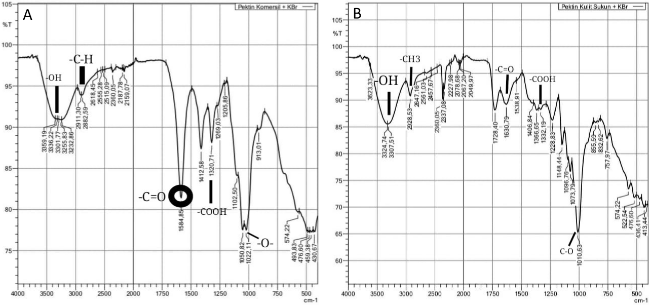
The functional groups of pectin isolated from breadfruit peel were characterized using Fourier Transform Infrared (FT-IR) spectroscopy. This analysis was conducted to confirm the chemical structure and identify the characteristic functional groups of the extracted pectin. The FT-IR spectrum of breadfruit peel pectin is presented in Figure 2. The spectral profile provides information on the presence of key functional groups associated with pectin, particularly those related to polysaccharide structures and esterified carboxyl groups, which are essential for its gel-forming and polymeric properties. The identification of these functional groups supports the successful isolation of pectin from breadfruit peel and confirms its suitability for use as a polymer matrix in floating bead formulations.
| No | Wavenumber range (cm-1) | Functional Group | ||
|---|---|---|---|---|
| Standar | Commercial Pectin | Sample Pectin | ||
| 1 | 3200-3600 | 3200-3500 | 3200-3500 | -OH |
| 2 | 2850-2950 | 2900-2950 | 2900-2950 | -CH3 |
| 3 | 1650-1750 | 1730-1750 | 1730-1750 | -C=O |
| 4 | 1600-1650 | 1600-1650 | 1600-1650 | -COOH |
| 5 | 1200-1000 | 1200-1000 | 1200-1000 | C-O (eter) |
The FT-IR spectral data indicate strong similarities between commercial pectin and the pectin extracted from breadfruit peel. Both spectra exhibited a broad absorption band in the region of 3200–3500 cm⁻¹, corresponding to O–H stretching vibrations associated with hydrogen-bonded hydroxyl groups from sugar units and bound water. This confirms the abundance of hydroxyl groups, which are characteristic of polysaccharide structures such as pectin. Absorption peaks observed at 2850–2950 cm⁻¹ represent C–H stretching vibrations of aliphatic –CH₃ groups derived from the monosaccharide backbone of pectin. The band detected in the range of 1650–1750 cm⁻¹ corresponds to C=O stretching of methyl-esterified galacturonic acid groups. The similarity in peak positions between the commercial and extracted samples suggests that the isolated pectin possesses a degree of esterification comparable to that of commercial pectin. Furthermore, absorption bands in the region of 1600–1650 cm⁻¹ indicate the presence of non-esterified galacturonic acid units, reflecting free COOH groups that play an important role in gel formation and ionic interactions. The fingerprint region at 1200–1000 cm⁻¹ corresponds to C–O–C and C–O stretching vibrations of ether linkages typical of polysaccharide structures. Overall, the identified functional groups including hydroxyl (O–H), aliphatic (C–H), ester carbonyl (C=O), carboxylic (–COOH), and ether linkages—confirm that the extracted material exhibits structural characteristics consistent with pectin. These findings demonstrate that the isolation process successfully produced pectin with functional group profiles comparable to commercial pectin (19). The FT-IR spectra of breadfruit peel-derived pectin and commercial pectin exhibited similar characteristic absorption bands associated with hydroxyl, carbonyl, and glycosidic functional groups. No substantial peak shifts or disappearance of characteristic bands were observed after drug incorporation. This finding suggests that amoxicillin was primarily incorporated into the floating bead matrix through physical entrapment rather than significant chemical interaction with the pectin polymer. Although slight differences in peak intensity were observed between commercial pectin and breadfruit peel-derived pectin, the major characteristic absorption bands appeared within similar wavenumber regions. These findings suggest that both materials possess comparable functional group compositions and potentially similar gel-forming capabilities. However, because degree of esterification and methoxyl content were not determined, definitive conclusions regarding differences in esterification characteristics could not be established.
Entrapment Efficiency Results
The entrapment efficiency of amoxicillin in all bead formulations is presented in Figure 3. This parameter reflects the ability of the polymer matrix to retain the drug within the bead structure during the gelation and curing processes. Variations in entrapment efficiency among the formulations are influenced by differences in pectin type and concentration, which affect matrix density, crosslinking behavior, and drug diffusion during bead formation. Higher polymer concentrations generally promote stronger gel networks, which can enhance drug retention by reducing drug leakage into the external medium during ionotropic gelation. A detailed comparison of the entrapment efficiency values for each formulation is illustrated in Figure 3.
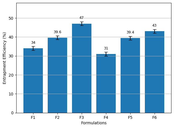
Drug entrapment is influenced by the surface area of the beads and the extent of crosslinking formed through the penetration of Ca²⁺ ions. Calcium ions interact with the carboxyl groups of pectin to form a three-dimensional “egg-box” network, which stabilizes the polymer matrix, retains the drug within the beads, and contributes to controlled drug release (20). The entrapment efficiency of amoxicillin in formulations F1, F2, and F3 (containing commercial pectin) ranged from 34% to 47%, while formulations F4, F5, and F6 (containing breadfruit peel–derived pectin) showed entrapment efficiencies between 31% and 43%. These differences were primarily influenced by the amount of pectin used in each formulation. Higher pectin concentrations resulted in increased entrapment efficiency due to the formation of a denser and more cohesive polymer network. At a lower pectin concentration (3.0%) in formulations F1 and F4, the polymer–drug mixture exhibited lower viscosity, producing smaller and more numerous droplets during bead formation. This increased the surface area exposed to the CaCl₂ solution, facilitating greater drug diffusion into the external medium and resulting in higher amounts of unentrapped drug. In contrast, formulations containing 3.50% pectin (F3 and F6) produced more viscous mixtures that generated larger and fewer droplets. This reduced the surface area available for drug loss during gelation, thereby decreasing the amount of free drug and improving entrapment efficiency (21). Although the entrapment efficiency values obtained in this study were moderate (31–47%), a concentration-dependent increase was observed in both commercial and breadfruit peel-derived pectin formulations. The relatively low entrapment efficiency may be attributed to the high aqueous solubility of amoxicillin, which facilitates drug diffusion into the external calcium chloride solution during ionotropic gelation. Further optimization of polymer concentration, crosslinking conditions, and drug-to-polymer ratio may improve drug retention within the polymer matrix.
Amoxicillin Release Test Results from Beads Preparation
In Vitro Drug Release from Floating Beads
The in vitro drug release study demonstrated that calcium ions (Ca²⁺) played a crucial role in enhancing the structural integrity of the pectin-based gel matrix, thereby prolonging the release of amoxicillin from the floating beads. The formation of a stronger gel network due to ionic crosslinking contributed to a sustained drug release behavior. The release mechanism of amoxicillin from the floating beads is governed by a combination of swelling, diffusion, and erosion processes, which collectively regulate the drug release profile. In an acidic medium, the repulsion between the carboxylic acid groups within the pectin structure is reduced, allowing the polymer chains to remain compact while gradually absorbing the surrounding medium. This condition facilitates controlled swelling of the beads, followed by diffusion of the drug through the hydrated gel layer. Additionally, matrix erosion contributes to the gradual release of the entrapped drug over time (15). The cumulative release profiles of amoxicillin from formulations F1–F6 are presented in Figure 4.
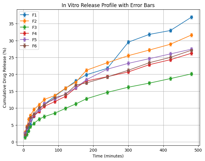
All formulations exhibited a sustained release pattern that met the established criteria for sustained-release systems. According to the criteria reported by Ref. (22), sustained-release dosage forms are generally expected to release approximately 10–25% of drug at 3 h, 15–30% at 6 h, and 20–40% at 8 h. These criteria were used in the present study as a comparative reference for evaluating sustained-release performance. The results showed that all floating bead formulations complied with these requirements, indicating effective control over drug release. In contrast, the conventional tablet formulation exhibited a rapid drug release, reaching 64.77% within 2 h, highlighting the advantage of the floating bead system in prolonging drug release. The drug release profiles of all floating bead formulations differed significantly from that of the conventional tablet formulation. One-way ANOVA followed by Tukey's HSD post hoc analysis demonstrated significant differences between the floating bead formulations and the conventional tablet formulation (p < 0.001). Pairwise comparisons further revealed significant differences among most formulation pairs, although several formulations exhibited statistically comparable release behavior. The significantly slower drug release observed in the floating bead formulations may be attributed to the formation of a hydrated and crosslinked pectin matrix, which acts as a diffusion barrier and prolongs drug release. This observation is consistent with the swelling behavior of the formulations, where matrix hydration contributed to sustained drug diffusion over time. Consequently, the floating bead formulations exhibited a slower and more controlled release pattern than the conventional tablet, suggesting that the pectin matrix effectively regulated drug diffusion throughout the dissolution period.
Release Kinetics Analysis of Amoxicillin from Floating Beads
The correlation coefficient (R²) values of amoxicillin release kinetics for all floating bead formulations are presented in Table 6.
| Formulation | Zero Order (R2) | First Order (R2) | Higuchi (R2) | Korsmeyer Peppas (R2) | n |
|---|---|---|---|---|---|
| F1 (Commercial Pectin 3.0%) | 0.991 | 0.873 | 0.995 | 0.989 | 0.623 |
| F2 (Commercial Pectin 3.25%) | 0.985 | 0.902 | 0.993 | 0.982 | 0.601 |
| F3 (Commercial Pectin 3.50%) | 0.978 | 0.910 | 0.989 | 0.975 | 0.588 |
| F4 (Breadfruit Peel Pectin 3.0%) | 0.992 | 0.884 | 0.996 | 0.990 | 0.645 |
| F5 (Breadfruit Peel Pectin 3.25%) | 0.989 | 0.895 | 0.994 | 0.987 | 0.632 |
| F6 (Breadfruit Peel Pectin 3.50%) | 0.987 | 0.901 | 0.993 | 0.986 | 0.618 |
The correlation coefficient (R²) values indicate that the Higuchi model exhibited the highest R² values for all formulations, suggesting that amoxicillin release followed the Higuchi diffusion model. This indicates that drug release was primarily controlled by diffusion through the polymeric matrix (8). In this system, the drug is dispersed within the matrix and gradually released through the pores of the polymer network over time (23). The zero-order model also showed relatively high R² values, indicating a tendency toward constant drug release. However, since the Higuchi model demonstrated higher R² values, diffusion remained the dominant release mechanism. Meanwhile, lower R² values in the first-order model indicate that drug release was not solely dependent on the remaining drug concentration within the system (24). Further analysis using the Korsmeyer–Peppas model showed that the diffusion exponent (n) values for all formulations ranged between 0.5 and 0.7. These values indicate a non-Fickian (anomalous) transport mechanism, which involves a combination of drug diffusion and polymer matrix relaxation processes such as swelling and erosion (23). This result suggests that amoxicillin release from the floating beads was not governed solely by diffusion but also influenced by the swelling behavior of the pectin matrix and gradual erosion of the polymer structure. The hydrophilic nature of pectin allows the beads to absorb simulated gastric fluid, forming a gel layer that controls drug diffusion. Simultaneously, matrix erosion contributes to sustained drug release over an extended period. The selection of the most appropriate release model in the present study was primarily based on correlation coefficient (R²) values, which is a commonly applied approach in preliminary release kinetic evaluations. Nevertheless, additional model validation approaches may provide a more comprehensive assessment of release behavior and should be considered in future investigations.
Limitations and Future Perspectives
Although the present study demonstrated the potential applicability of breadfruit peel-derived pectin as a matrix-forming polymer for gastroretentive floating beads, several limitations should be acknowledged. In addition, bead diameter and weight measurements were performed using five randomly selected beads per replicate batch. Although this sampling approach provided preliminary characterization of bead morphology, a larger sample size would provide a more representative assessment of bead size distribution and variability. Therefore, future studies should include a greater number of beads for dimensional and weight analysis. The study did not include commercially available sustained-release benchmark formulations or dissolution similarity factor analysis. In addition, further physicochemical characterization of the extracted pectin, including degree of esterification, methoxyl content, galacturonic acid content, moisture content, and viscosity determination, would provide a more comprehensive understanding of its pharmaceutical properties and their influence on bead formation, drug entrapment efficiency, swelling behavior, and drug release characteristics. Future studies should address these aspects and explore formulation optimization strategies to improve drug entrapment efficiency and overall formulation performance.
Conclusion
Amoxicillin gastroretentive floating beads were successfully formulated using both breadfruit peel-derived pectin and commercial pectin through ionotropic gelation. The formulated beads exhibited satisfactory floating properties, swelling behavior, and sustained-release characteristics. Increasing pectin concentration generally improved drug entrapment efficiency and prolonged drug release. The release profiles were most closely described by the Higuchi model, suggesting that diffusion played an important role in the drug release mechanism under the experimental conditions investigated. The findings suggest that breadfruit peel-derived pectin has potential as an alternative natural polymer for gastroretentive floating bead formulations. However, variations in formulation performance, particularly entrapment efficiency, indicate that further optimization is required. In addition, the present study was limited to in vitro evaluation and did not include comprehensive physicochemical characterization, long-term stability testing, toxicity evaluation, or in vivo gastric retention studies. Therefore, further investigations are necessary to confirm the pharmaceutical applicability of breadfruit peel-derived pectin and to evaluate its performance under physiological conditions.
Abbreviations
GRDDS = Gastroretentive Drug Delivery System; FT-IR = Fourier Transform Infrared; NaHCO₃ = Sodium Bicarbonate; CaCl₂ = Calcium Chloride; SD = Standard Deviation; ANOVA = Analysis of Variance; USP = United States Pharmacopeia; API = Active Pharmaceutical Ingredient.
Declarations
Conflict of Interest
The authors declare no conflict of interest.
Data Availability
The datasets generated and/or analyzed during the current study are included in this article and are available from the corresponding author upon reasonable request
Ethics Statement
Not applicable.
Funding Information
This work received no external funding.
References
- Moriya G, Mazumder R, Padhi S, Mishra R. Gastrorententive hydrogels responsive to external stimuli for novel drug delivery. Int J App Pharm. 2024;6(4):1-14. doi: https://doi.org/10.22159/ijap.2024v16i4.51051
- Raeisi A, Farjadian F. Commercial hydrogel product for drug delivery based on route of administration. Front. Chem. 2024;12:1336717. doi: https://doi.org/10.3389/fchem.2024.1336717
- Liu Y, Huang J, Li S, Li Z, Chen C, Qu G, et al. Advancements in hydrogel-based drug delivery systems for the treatment of inflammatory bowel disease: a review. Biomater. Sci. 2024;12(4):837-862. doi: https://doi.org/10.1039/d3bm01645e
- Sabbagh F, Zargarian SS, Kosik-Kozioł A, Nakielski P, Pierini F. Hydrogel-based ocular drug delivery systems. J. Mater. Chem. B. 2025;13(46):14982-15006. doi: https://doi.org/10.1039/d5tb01575h
- Zainol N, Zulfikree HS, Nor NFA, Yaakob H, Zaidel DNA. Characterization of Artocarpus altilis powder as a potential health supplement ingredient. Malays J Anal Sci. 2023;27(5):1147–59.
- Kedir WM, Deresa EM, Diriba TF. Pharmaceutical and drug delivery applications of pectin and its modified nanocomposites. Heliyon. 2022;8(9):e10654. doi: https://doi.org/10.1016/j.heliyon.2022.e10654
- Candrika C, Fauzi ZPA, Puteri CIA, Damanik FS, Utami DS, Hartini PT. Senggani Leaf Extract-Based Gel Facial Wash: A Herbal Innovation for Anti-Acne Therapy Targeting Cutibacterium acnes. Jps. 2026;9(1):783-796. doi: https://doi.org/10.36490/journal-jps.com.v9i1.1454
- Muniyandy S. Pectin beads in drug delivery: extraction, formulation, and pharmaceutical applications. Int J App Pharm. 2025;17(1):64-73. doi: https://doi.org/10.22159/ijap.2025v17i5.54903
- Sen S, Sinha R, Giri AH, Kumari P, Kumbhar AP, Ranjan OP. Nanomaterials and Nanocomposites from Wastes for Drug Delivery and Advanced Therapy: A Review. BioNanoSci. 2025;15(1). doi: https://doi.org/10.1007/s12668-024-01769-3
- Rajmane A, Trivedi R, Nandgude T. A Novel approach to Enhance Gastric Retention for better Therapeutic Activity: Gastro Retentive Drug Delivery System. Rjpt. 2022:3324-3330. doi: https://doi.org/10.52711/0974-360x.2022.00556
- Samir K. S, D. Barhate S. Formulation Development and Evaluation of Sustained Release Floating Beads of Aspirin. Rjpdft. 2025:97-101. doi: https://doi.org/10.52711/0975-4377.2025.00014
- Almasri R, Swed A, Alali H. Preparation and Characterization of Hydrogel Beads for Controlled Release of Amoxicillin. Jordan j. pharm. sci. 2022;15(4):523-535. doi: https://doi.org/10.35516/jjps.v15i4.675
- Ghasemzadeh M, Gozalzadeh S, Sirousazar M, Kheiri F. Amoxicillin-loaded bionanocomposite hydrogels: swelling, dehydration, and in vitro drug release kinetics and mechanism. Journal of Biomaterials Science, Polymer Edition. 2023;35(4):463-481. doi: https://doi.org/10.1080/09205063.2023.2295058
- Sipos B, Benei M, Katona G, Csóka I. Optimization and Characterization of Sodium Alginate Beads Providing Extended Release for Antidiabetic Drugs. Molecules. 2023;28(19):6980. doi: https://doi.org/10.3390/molecules28196980
- Maulina AR, Irdawati I. Characterization of Immobilized Cell Alginate Beads of Mudiak Sapan (MS) Isolates Producing Bioelectricity. Jbt. 2025;25(2):1414-1418. doi: https://doi.org/10.29303/jbt.v25i2.8826
- Siraj EA, Mulualem Y, Molla F, Yayehrad AT, Belete A. Formulation optimization of furosemide floating-bioadhesive matrix tablets using waste-derived Citrus aurantifolia peel pectin as a polymer. Sci Rep. 2025;15(1). doi: https://doi.org/10.1038/s41598-025-95732-1
- Wahba MI. Boosting the stability of β-galactosidase immobilized onto soy-protein isolate-glutaraldehyde-functionalized carrageenan beads. 3 Biotech. 2023;13(1). doi: https://doi.org/10.1007/s13205-022-03446-2
- Dhiman S, Philip N, Gurjeet Singh T, Babbar R, Garg N, Diwan V, et al. An Insight on Novel Approaches & Perspectives for Gastro-Retentive Drug Delivery Systems. Cdd. 2023;20(6):708-729. doi: https://doi.org/10.2174/1567201819666220819200236
- Askarizadeh M, Esfandiari N, Honarvar B, Sajadian SA, Azdarpour A. Kinetic Modeling to Explain the Release of Medicine from Drug Delivery Systems. ChemBioEng Reviews. 2023;10(6):1006-1049. doi: https://doi.org/10.1002/cben.202300027
- Nesic A, Meseldzija S, Onjia A, Cabrera-Barjas G. Impact of Crosslinking on the Characteristics of Pectin Monolith Cryogels. Polymers. 2022;14(23):5252. doi: https://doi.org/10.3390/polym14235252
- Manna S, Aziz A, Das S, Bhowmik M. Ionically Cross-linked Pectin-alginate Hydrogel Beads for Prolonged Release of Glibenclamide. Int J. Pharm. Investigation. 2022;12(4):475-478. doi: https://doi.org/10.5530/ijpi.2022.4.81
- Hsu YT, Kao CY, Ho MH, Lee SP. To control floating drug delivery system in a simulated gastric environment by adjusting the Shell layer formulation. Biomater Res. 2021;25(1). doi: https://doi.org/10.1186/s40824-021-00234-6
- Krishnarajan D, Reddy CM, Kanikanti S, Kumar NS, Purushothaman M. Formulation and evaluation of sustained release matrix tablets Of Levofloxacin using natural polymer. Pharmacophore. 2013;4(5–2013):146–57.
- Siswanto A, Fudholi A, Nugroho AK, Martono S. In vitro release modeling of aspirin floating tablets using ddsolver. Indonesian J. Pharm. 2015;26(2):94. doi: https://doi.org/10.14499/indonesianjpharm26iss2pp94