RESEARCH ARTICLE
Ancestral Heritage Toward Health Innovation: A Study of the Antibacterial Activity of Betel Leaf (Piper betle Linn.) Extract from the Betel-Chewing Tradition Against Oral Pathogenic Bacteria
Academic Editor: Garnadi Jafar
Sciences of Pharmacy|Vol. 5, Issue 3, pp. 261-272 (2026)
CC BY 4.0-2026 Authors
Views
Downloads
Shares

Received
Apr 18, 2026Revised
May 25, 2026Accepted
Jun 18, 2026Published
Jul 10, 2026
Abstract
Traditionally, menginang (a mixture of betel leaf, areca nut, gambir, and lime) has been used as a natural antibacterial agent. This study aimed to compare the antibacterial activity of single betel leaf extract and menginang extract against four oral pathogenic bacteria in vitro. This study compared the antibacterial activity of traditional menginang extract and single betel leaf extract against multiple oral pathogenic bacteria to evaluate their potential synergistic antibacterial effects. The experimental method used a disk diffusion technique at three concentrations (12.5%, 25%, and 50%), and the data were analyzed using one-way ANOVA and Bonferroni post hoc tests. Chlorhexidine 0.2% was used as the positive control, while distilled water was used as the negative control. The 50% menginang extract showed the strongest antibacterial activity against all tested bacteria, with inhibition zones ranging from 22.76 ± 1.38 mm to 28.22 ± 0.14 mm, and the differences were statistically significant (p < 0.05). The inhibition zones produced by the 50% menginang extract were close to those of 0.2% chlorhexidine. The superiority antibacterial activity of menginang extract may be associated with synergistic interactions among alkaloids, phenolics, flavonoids, tannins, saponins, triterpenoids, and steroids identified during phytochemical screening. The menginang extract has high potential to be developed as an innovative natural product for oral health.
Introduction
The mouth is home to a variety of microorganisms, including bacteria, which are naturally present on its surface (1). Oral pathogens can cause infections in the oral cavity and contribute to the development of dental caries, gingivitis, periodontitis. In particular, certain species are clinically important because they play specific roles in biofilm formation and periodontal destruction. The Ministry of Health of the Republic of Indonesia reports that 25.9% of the Indonesian population has dental health issues, and 57.6% of Indonesians have dental problems. This shows that oral and dental diseases remain a major public health problem in Indonesia (2).
The bacterium Streptococcus mutans is often the cause of caries, as it is a major acid-producing and acid-tolerant bacterium involved in enamel demineralization. Oral and dental health problems are still very common in Indonesia. Based on the 2023 Indonesian Health Survey (SKI), as many as 56.9% of the population aged over three years have dental and oral problems, with 88% of them suffering from caries or cavities. The anaerobic gram-negative bacteria Porphyromonas gingivalis plays a role in the development of periodontitis through its contribution to subgingival dysbiosis and periodontal tissue damage. Prevalence of periodontitis in Indonesia reached 74.1% in the population aged 15 years and above (women: 74.7%, men: 73.2%). The prevalence of oral health problems increased from 25.9% (2013) to 57.6% (2018). Only 6.2% of Indonesians brush their teeth with the correct time and technique as recommended (morning after breakfast and night before bed), while 58.5% do not replace their toothbrushes every three months. Poor oral hygiene facilitates the buildup of plaque and calculus and contributes to the severity of gum disease. Among 5-year-old children, 91.2% and 12-year-old children, 94.1% have never been taken to a dental health professional for treatment (3). Aggregatibacter actinomycetemcomitans is a gram-negative facultative anaerobic bacterium that produces several virulence factors and has high genetic diversity. Fusobacterium nucleatum is also clinically important because it acts as a bridge organism in oral biofilms and supports the colonization of other periodontal pathogens (4).
A. actinomycetemcomitans is capable of traversing epithelial barriers and perturbing both oral and gut microbial communities, with potential consequences for systemic health. Its invasive behavior and toxin production promote local inflammation and can facilitate translocation into deeper tissues and the bloodstream. Systemically, A. actinomycetemcomitans has been implicated in conditions such as rheumatoid arthritis and infective endocarditis, and it may contribute to carcinogenesis through induction of pro‑inflammatory cytokines (for example, IL‑1β and TNF), which create a microenvironment conducive to chronic inflammation and tissue damage (5).
P. gingivalis is a keystone anaerobic pathogen in periodontitis that employs multiple strategies to invade host cells and evade innate and adaptive immune responses. P. gingivalis secretes proteolytic gingipains and other enzymes that degrade host proteins, dysregulate immune signaling, and manipulate complement and toll‑like receptor pathways. Notably, P. gingivalis can induce protein citrullination a posttranslational modification linked to autoimmunity providing a mechanistic link between periodontal infection and systemic diseases such as rheumatoid arthritis; associations have also been reported between P. gingivalis and systemic inflammation, atherosclerosis, and oral mucositis (6).
F. nucleatum is a Gram‑negative anaerobe implicated in both periodontal pathology and a range of systemic conditions, most prominently colorectal cancer. F. nucleatum expresses adhesins and other virulence factors that promote robust biofilm formation, modulate host immune responses, and disrupt epithelial barrier integrity, thereby facilitating local invasion and systemic dissemination. Clinical and molecular studies have frequently detected Fn not only in periodontal pockets but also at distant sites such as atherosclerotic plaques and colorectal tumors supporting the hypothesis that hematogenous spread of oral microbes can contribute to chronic inflammation and disease processes beyond the oral cavity (7).
S. mutans exhibits exceptional capacity to form robust biofilms on dental surfaces, which underpins its cariogenic potential. This phenotype is largely driven by glucosyltransferases (gtfB, gtfC, gtfD), which catalyze the production of extracellular polysaccharides (EPS). The resultant EPS matrix promotes initial adherence, interbacterial cohesion, and structural stability of the biofilm while conferring protection against acid, oxidative stress, and antimicrobial agents (8).
Menginang is a traditional practice of chewing betel leaf mixed with lime, gambir, and A. nut. This practice has been preserved across generations and remains an important part of local culture. In the menginang tradition, betel leaves are believed to be beneficial for dental health, possess strong antifungal properties, and are capable of inhibiting the growth of certain types of bacteria. Because of this, menginang has attracted interest as a potential natural antibacterial source (9).
Betel or A. nut has become a customary practice in the daily lives of the people, particularly in Banjarmasin, South Kalimantan. Menginang is the practice of chewing a mixture of betel leaves, A. nuts, lime, and gambir. In the menginang tradition, betel leaves are believed to be beneficial for dental health, possess strong antifungal properties, and are capable of inhibiting the growth of certain types of bacteria (10).
Previous studies have reported the antibacterial activity of Piper betle, Areca catechu, and gambir extracts against several pathogenic microorganisms, including oral bacteria related to caries and periodontal disease. However, studies evaluating the synergistic antibacterial activity of the traditional menginang combination against multiple oral pathogenic bacteria remain limited. In particular, direct comparative studies between single betel leaf extract and traditional menginang extract are still scarce. Therefore, this study was conducted to evaluate and compare the antibacterial effectiveness of both extracts against clinically important oral pathogens associated with dental caries and periodontal disease.
Previous studies have demonstrated that the combination of P. betle, A. catechu, Uncaria gambir, and lime exhibits synergistic antibacterial effects. Lime contributes by increasing the alkalinity of the mixture, which may enhance the release and activity of bioactive compounds from the plant materials and create unfavorable conditions for bacterial growth. The stronger antibacterial activity of the menginang extract may be attributed to synergistic interactions among bioactive compounds derived from P. betle, U. gambir, and A. catechu. Phenolic compounds, hydroxychavicol, catechins, tannins, and alkaloids collectively disrupt bacterial membrane integrity, interfere with protein synthesis, and alter membrane permeability, thereby enhancing antibacterial efficacy against oral pathogenic bacteria. These combined mechanisms enhance antibacterial activity against oral pathogenic bacteria, including S. mutans, P. gingivalis, and A. actinomycetemcomitans (13, 14, 16).
The objectives of this study were: 1). to analyze the effectiveness and optimal concentration of betel leaf (P. betle L.) extract in inhibiting the growth of oral pathogenic bacteria, including A. actinomycetemcomitans, S. mutans, P. gingivalis, and F. nucleatum in vitro; 2). to assess the effectiveness and optimal concentration of extracts derived from the traditional menginang formulation in inhibiting the growth of oral pathogenic bacteria, including A. actinomycetemcomitans, S. mutans, P. gingivalis, and F. nucleatum in vitro; and 3). to compare the effectiveness between betel leaf extract and menginang plant extracts in producing the most optimal inhibition zone diameter against the growth of oral pathogenic bacteria.
This study aimed to provide empirical data on the effectiveness of betel leaves used in the betel-chewing tradition, as compared to betel leaves alone, in inhibiting the growth of pathogenic bacteria in the oral cavity. The findings of this study may provide scientific evidence for the development of natural oral health products and support the preservation of the traditional menginang practice.
Methodology
Time and Place
This study was conducted from July to September 2025 at the Microbiology Laboratory and the Biology Laboratory, Faculty of Pharmacy, Muhammad Arsyad Al Banjari Islamic University of Kalimantan. During this period, the study focused on experimental procedures, including sample preparation and laboratory analysis, while data processing and manuscript preparation were carried out separately after the experimental phase
Materials
The materials used in this study were betel leaves (P. betle Linn.), A. nut (A. catechu L.), gambir (U. gambir (Hunter) Roxb.) whose authenticity had been verified, betel lime, nutrient agar (NA), nutrient broth (NB), test bacteria (A. actinomycetemcomitans, S. mutans, P. gingivalis, and F. nucleatum), disc paper, chlorhexidine, 70% ethanol, distilled water, NaCl solution, and Mueller-Hinton agar (MHA).
Research Method
This study was experimental in nature, employing a laboratory test model using the disc diffusion method to determine antibacterial activity. The independent variable was the concentration of betel leaf and menginang extracts at three concentration levels (12.5%, 25%, and 50%), while the dependent variable was the growth of pathogenic bacteria, measured by the diameter of the inhibition zone. The selected concentrations (12.5%, 25%, and 50%) were determined based on previous studies evaluating the antibacterial activity of P. betle L. extract against oral pathogenic bacteria using graded concentration levels, as well as preliminary screening tests conducted to observe concentration-dependent antibacterial effects (11). Control variables included incubation temperature (37°C), incubation time (24 hours), and positive-negative controls for result validation.
Phytochemical Screening
Phytochemical screening is the preliminary stage of phytochemical research aimed at providing an overview of the classes of compounds contained in the plant under study. The discovery of new drugs is of great importance to every local government in the country at this time. One natural resource with the potential to contain bioactive compounds that can function as medicines is biological resources, particularly plants belonging to the Piperaceae family. Therefore, to increase the utility of Piperaceae family members domestically, further and continuous research is needed to identify bioactive compounds that can subsequently be produced as medicines in the pharmaceutical industry (12).
Preparation of Betel Leaf Extract and Menginang Extract
The ingredients for menginang (betel leaf, A. nut, lime, and gambir) were thoroughly washed, dried, and ground using a grinder. A total of 250 grams of betel leaf powder and 62.5 grams of each menginang ingredient were weighed and extracted using 70% ethanol via the maceration method, with the soaking process conducted for 2–3 days, followed by evaporation using a rotary evaporator (13). Betel leaf extract and Menginang extract were prepared at three concentration levels: 12.5%, 25%, and 50%. Concentrations were prepared by gradual dilution (14). The extraction process used a solvent-to-sample ratio of 10: 1 (v/w). The menginang formulation consisted of betel leaf, A. nut, gambir, and lime in a ratio of 1: 1: 1: 1. The extraction yield was calculated using the following formula:
Bacterial Rejuvenation and Preparation of Bacterial Suspensions
Bacteria were collected using a sterile inoculation loop and streaked onto the surface of Nutrient Agar (NA). The streaked agar was then incubated at 37°C for 24 hours. A loopful of rejuvenated bacterial culture from Nutrient Agar (NA) was inoculated into 5 mL of Nutrient Broth (NB) and incubated at 37°C for 24 hours. The bacterial suspension turbidity was then adjusted to match the 0.5 McFarland standard, equivalent to approximately 1.5 × 10⁸ CFU/mL, to standardize the bacterial inoculum used in the antibacterial assay (14).
Antibacterial Efficacy via the Disc Diffusion Method
200 μl of bacterial suspension was added to 20 ml of Mueller Hinton Agar (MHA) medium, poured into a petri dish, and allowed to solidify. 10 μl of the sample solution was applied to a paper disc. The discs containing the sample were then placed on the surface of the agar medium. After incubation for 24 hours at 37°C, the clear areas (inhibition zones) formed around the discs were observed and their sizes recorded (14). Chlorhexidine 0.2% was used as the positive control, while distilled water was used as the negative control.
Data Analysis
The determination of bacterial inhibition test results using the disk diffusion method is based on the presence of an inhibition zone around the paper disk. After the average inhibition zone is obtained, the classification of inhibitory ability is determined according to Table 1. The data were statistically analyzed using a normality test, followed by a One-Way ANOVA and a Bonferroni post hoc test (14). All antibacterial assays were performed in triplicate to ensure data reliability and reproducibility. All microbiological procedures were carried out following laboratory biosafety guidelines for handling pathogenic bacteria. This study did not involve human or animal subjects; therefore, ethical clearance was not required.
| Growth Inhibition Responses | Inhibition Zone Diameter |
|---|---|
| Strong | > 20 mm |
| Moderate | 16-20 mm |
| Weak | 10-15 mm |
| None | < 10 mm |
Results and Discussion
Plant Determination
Betel leaves, gambir, and A. nuts were obtained from Hulu Sungai Tengah Regency, South Kalimantan, and identified at the Laboratory of the Faculty of Mathematics and Natural Sciences (FMIPA) at Lambung Mangkurat University in Banjarbaru. Based on certificate number: 163/LB. LABDASAR/VIII/2025, the samples used were confirmed to be P. betle Linn. Meanwhile, regarding gambir, based on certificate number: 195/LB. LABDASAR/IX/2025, the samples used were confirmed to be the plant U. gambir (Hunter) Roxb. Regarding A. nut, based on certificate number: 216/LB. LABDASAR/IX/2025, the samples used were confirmed to be the plant A. catechu L.
Phytochemical Screening
Phytochemical screening confirmed the presence of major bioactive compounds in betel leaf (P. betle L.) and menginang extracts, as presented in Table 2. Betel leaf extract tested positive for alkaloids (cream with Mayer, orange with Dragendorff, brown with Wagner), phenolics (bluish green with FeCl₃), flavonoids (orange-red with Shinoda/Mg-HCl), tannins (blackish green with FeCl₃), saponins (stable foam > 10 minutes), triterpenoids (orange Liebermann-Burchard), and steroids (blue/green Liebermann-Burchard). The menginang extract showed similar results: alkaloids (Mayer’s cream, Dragendorff’s orange, Wagner’s brown), phenolics (bluish-green with FeCl₃), flavonoids (Shinoda’s orange-red), tannins (blackish-green with FeCl₃), saponins (stable foam), and triterpenoids (Liebermann-Burchard orange). The diversity of these secondary metabolites in both extracts indicates strong antimicrobial, anti-inflammatory, and pharmacological potential (15).
| Phytochemical Compounds | Reagents / Methods | Observation (Positive Indicators) | Betel Leaf (P. betle) | Menginang |
|---|---|---|---|---|
| Alkaloids | Mayer’s Test | Creamy precipitate | (+) | (+) |
| Dragendorff’s Test | Orange precipitate | (+) | (+) | |
| Wagner’s Test | Brown precipitate | (+) | (+) | |
| Phenolics | FeCl₃ Test | Bluish-green coloration | (+) | (+) |
| Flavonoids | Shinoda (Mg + HCl) | Orange-red coloration | (+) | (+) |
| Tannins | FeCl₃ Test | Blackish-green coloration | (+) | (+) |
| Saponins | Froth Test | Stable foam (> 10 minutes) | (+) | (+) |
| Triterpenoids | Liebermann-Burchard | Orange coloration | (+) | (+) |
| Steroids | Liebermann-Burchard | Blue or Green coloration | (+) | (+) |
Phytochemical screening was conducted as a preliminary step to qualitatively detect the presence of secondary metabolites in menginang and betel leaf (P. betle L.) extracts. Analysis of the betel leaf extracts indicated the presence of alkaloids, phenolics, flavonoids, steroids, tannins, saponins, and triterpenoids in both extracts. Meanwhile, Menginang extract contained alkaloids, phenolics, saponins, triterpenoids, tannins, steroids, and flavonoids. The diversity of these bioactive compounds indicates strong biological potential, particularly in antimicrobial activity. The antibacterial activity observed in this study may be associated with the bioactive compounds detected during phytochemical screening, including alkaloids, phenolics, flavonoids, tannins, saponins, triterpenoids, and steroids. Previous studies have suggested that specific constituents such as hydroxychavicol, catechins, condensed tannins, and arecoline may contribute to these antibacterial effects.
The diversity of natural bioactive compounds in betel leaves, A. nuts, and gambir as components of the traditional betel-chewing mixture indicates that the antibacterial activity against these four species of oral pathogenic bacteria is supported by the synergy between phenolics, flavonoids, tannins, alkaloids, and saponins, which have been shown to exert antibacterial effects by disrupting bacterial membrane integrity, inducing leakage of intracellular components, inhibiting bacterial enzymes, and interfering with protein synthesis in both Gram-positive and Gram-negative bacteria in several previous studies, with the main differences lying in the concentrations and combinations of the raw materials used (16).
Betel Leaf and Menginang Extracts
The betel leaves obtained are dark green in color and have a distinctive odor, while the menginang is dark brown and has an aromatic odor characteristic of the menginang ingredients, as shown in Figure 1.

Menginang extract from 250 grams of crude drug powder yielded 25.7 grams of concentrated extract with a yield of 10.28% (> 10%, meets requirements), while betel leaves from 250 grams of crude drug powder yielded 28 grams of concentrated extract with a yield of 11.2% (> 10%, meets requirements). A low yield reflects suboptimal extraction efficiency, resulting in a small amount of extract obtained (17). The 11.2% yield of concentrated extract from betel leaf crude powder obtained in this study is relatively lower compared to the results of betel leaf essential oil extraction reported in other studies, where the essential oil yield can reach approximately 20.76 g/kg or 2% dry weight.
This difference can be explained by the fact that concentrated extracts contain a variety of bioactive compounds, whereas essential oil extraction focuses specifically on volatile compounds using different extraction methods, such as steam distillation. Additionally, other studies optimizing extraction methods using technologies such as cold plasma or ultrasonic- assisted extraction have also demonstrated increased yields and bioactive compound content from betel leaves and other herbal plants, underscoring the importance of appropriate extraction methods to maximize yields (18).
For betel leaf crude extract, specific literature data is indeed still limited; however, the 10.28% yield obtained falls within the range commonly found in the extraction of crude extracts from other herbal plants, which typically ranges from 8–15%. This indicates that the extraction process carried out was sufficiently optimal to obtain a concentrated extract containing active ingredients in adequate concentrations. Therefore, the results of this study are consistent with existing literature and provide a strong scientific basis for the development of products based on menginang and betel leaf extracts.
Antibacterial Efficacy Using the Disc Diffusion Method
Bacterial growth inhibition tests were conducted in triplicate across three concentration series of menginang and betel leaf extracts (50%, 25%, 12.5%), a positive control (0.2% chlorhexidine), and a negative control (distilled water) using the disk diffusion method. Inhibitory activity was indicated by inhibition zones around the paper disks, which were measured and averaged for each treatment. The representative inhibition zones at the 50% concentration are shown in Figure 2. The average inhibition zone values for each treatment are presented in Table 3.
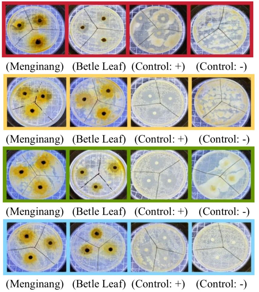
A recent study confirmed the potential of betel leaf (P. betle L.) in the disc diffusion method, where the green nano-extract produced a larger inhibition zone (up to 18.67 mm) against S. mutans compared to the micro-sized extract, thanks to the more efficient release of active compounds. Similarly, A. nut (A. catechu) extract demonstrated inhibitory activity against oral bacteria such as S. aureus with a clearly significant zone in the agar disc diffusion test, supporting the synergy in menginang mixtures for the development of natural oral health products (19).
| Bacteria | Concentration | Mean (mm) ± SD | Inhibition Response |
|---|---|---|---|
| P. gingivalis | 50% (Menginang) | 24.99 ± 3.21 | Strong |
| 25% (Menginang) | 22.95 ± 3.28 | Strong | |
| 12.5% (Menginang) | 22.68 ± 3.12 | Strong | |
| 50% (Betel) | 21.07 ± 2.40 | Strong | |
| 25% (Betel) | 15.63 ± 1.92 | Weak | |
| 12.5% (Betel) | 10.86 ± 1.64 | Weak | |
| (+) Chlorhexidine 0.2% | 27.98 ± 1.15 | Strong | |
| (-) Distilled water | 0 | None | |
| F. nucleatum | 50% (Menginang) | 28.22 ± 0.14 | Strong |
| 25% (Menginang) | 27.30 ± 0.22 | Strong | |
| 12.5% (Menginang) | 25.46 ± 1.83 | Strong | |
| 50% (Betel) | 17.19 ± 1.52 | Moderate | |
| 25% (Betel) | 15.38 ± 1.52 | Weak | |
| 12.5% (Betel) | 13.87 ± 0.94 | Weak | |
| (+) Chlorhexidine 0.2% | 27.78 ± 0.84 | Strong | |
| (-) Distilled water | 0 | None | |
| A. actinomycetemcomitans | 50% (Menginang) | 25.28 ± 2.03 | Strong |
| 25% (Menginang) | 22.54 ± 2.41 | Strong | |
| 12.5% (Menginang) | 16.07 ± 6.47 | Moderate | |
| 50% (Betel) | 17.75 ± 2.08 | Moderate | |
| 25% (Betel) | 16.13 ± 0.90 | Moderate | |
| 12.5% (Betel) | 12.12 ± 1.12 | Weak | |
| (+) Chlorhexidine 0.2% | 29.5 ± 0.00 | Strong | |
| (-) Distilled water | 0 | None | |
| S. mutans | 50% (Menginang) | 22.76 ± 1.38 | Strong |
| 25% (Menginang) | 21.09 ± 1.66 | Strong | |
| 12.5% (Menginang) | 20.68 ± 1.67 | Strong | |
| 50% (Betel) | 19.86 ± 1.91 | Moderate | |
| 25% (Betel) | 18.21 ± 1.69 | Moderate | |
| 12.5% (Betel) | 12.84 ± 1.22 | Weak | |
| (+) Chlorhexidine 0.2% | 28.75 ± 1.20 | Strong | |
| (-) Distilled water | 0 | None |
The results of the antibacterial testing of Menginang extract and betel leaf extract (P. betle Linn.) in Table 3. show that Menginang extract exhibits stronger and more consistent antibacterial activity against the four major oral pathogenic bacteria compared to betel leaf extract alone. Gram-positive bacteria are generally more susceptible to phenolic compounds than Gram-negative bacteria because their simpler peptidoglycan-rich cell wall lacks the outer membrane that acts as a permeability barrier in Gram-negative bacteria. This structural difference may explain the stronger inhibition observed against S. mutans compared with several Gram-negative oral pathogens tested in the present study. In addition, the multi-target interactions among phenolics, flavonoids, tannins, catechins, and alkaloids in the menginang extract may further enhance membrane disruption, enzyme inhibition, and bacterial growth suppression (20). The higher susceptibility of S. mutans may also be associated with the absence of an outer lipopolysaccharide membrane, allowing phenolic and flavonoid compounds to diffuse more easily into the bacterial cell. In contrast, Gram-negative bacteria such as P. gingivalis and A. actinomycetemcomitans possess an additional outer membrane barrier that can reduce the penetration of antibacterial compounds.
This difference in susceptibility may also be influenced by variations in bacterial cell wall composition and metabolic characteristics. Gram-positive bacteria possess a thick peptidoglycan layer without an outer lipopolysaccharide membrane, allowing antibacterial compounds such as hydroxychavicol, flavonoids, and tannins to diffuse more effectively into the bacterial cell. In contrast, Gram-negative bacteria have an additional outer membrane that acts as a permeability barrier and may reduce the penetration of bioactive compounds. Furthermore, the synergistic interaction among phenolic compounds, alkaloids, catechins, and tannins in the menginang extract may enhance membrane disruption, protein denaturation, and enzyme inhibition, thereby increasing antibacterial efficacy against oral pathogenic bacteria. These findings suggest that the antibacterial activity of the menginang extract is not solely concentration-dependent but may also result from synergistic interactions among its phytochemical constituents. The antibacterial activity observed in this study may be attributed to the presence of potent bioactive compounds in P. betle, such as hydroxychavicol, which exhibits significant antibacterial activity against various oral pathogens, including S. mutans.
Extracts of P. species, principally hydroxychavicol as the major bioactive constituent, exhibited strong antibiofilm activity against S. mutans by inhibiting biofilm formation and reducing preformed biofilms, actions that correlate with disruption of microbial membrane integrity and inhibition of insoluble glucan synthesis (21). The study also confirms that P. betle extract exhibits dose- dependent antibacterial effects and may serve as a promising candidate for future natural oral antibacterial development in the management of oral diseases, reinforcing the results of the Menginang extract tests, which showed large inhibition zones approaching the efficacy of chlorhexidine (19).
P. betle contains bioactive constituents such as acetyleugenol and hydroxychavicol that inhibit bacterial virulence factors. For example, acetyleugenol has been shown to bind Sortase A (SrtA), a critical enzyme mediating bacterial adhesion and pathogenesis, thereby attenuating virulence. Hydroxychavicol targets cell division proteins including FtsZ and FtsA, disrupting cytokinesis and inhibiting bacterial replication and growth. Hydroxychavicol exhibited antibacterial activity against multidrug‑resistant avian pathogenic Escherichia coli (APEC) with MIC and MBC values in the range of 0.25–1.0 mg·mL−1, and produced a rapid bactericidal effect (≥3‑log reduction in viable counts within 4 h at 4×MIC) (22).
Additionally, the strong antibacterial activity of the menginang extract may result from synergistic interactions among its phytochemical constituents. Extracts of P. species have been shown to modulate the activity of conventional antibiotics, enhancing their efficacy against resistant bacterial strains; this suggests that combining P. extracts with other agents, such as A. catechu, may potentiate antibacterial effects (23). Phenolic compounds and hydroxychavicol from P. betle may disrupt bacterial membrane integrity and increase membrane permeability. Catechins and condensed tannins from U. gambir may inhibit bacterial enzymes and interfere with protein synthesis, while alkaloids such as arecoline from A. catechu may alter bacterial metabolic pathways. The combined multi-target mechanisms of these bioactive compounds likely enhance antibacterial efficacy against oral pathogenic bacteria. Scanning electron microscopy (SEM) revealed morphological alterations in bacterial cells exposed to P. betle extract, which supports the hypothesis that disruption of membrane integrity is a primary antibacterial mechanism (24). These findings support previous ethnopharmacological studies reporting synergistic antibacterial effects of traditional herbal combinations and suggest that menginang extract has potential as a natural antibacterial agent for oral health applications (25). These compounds interfere with protein synthesis by binding to key enzymes of the translation process and causing changes in gene expression associated with protein regulation (26).
These findings are consistent with previous ethnopharmacological studies reporting that the combination of P. betle and A. catechu exhibits synergistic antibacterial effects against oral pathogens. In previous ethnopharmacological studies, the antibacterial activity of P. betle-A. catechu combinations was generally evaluated against a limited number of oral microorganisms and often produced moderate inhibition zones. The current findings demonstrated strong inhibitory activity against four clinically relevant oral pathogens, with inhibition zones reaching 28.22 mm at the 50% menginang concentration. Furthermore, the observed antibacterial activity approached that of 0.2% chlorhexidine, suggesting that the traditional menginang formulation may provide a stronger synergistic effect than previously reported single-plant or dual-component preparations. Previous studies have shown that Gram-positive bacteria such as S. mutans are generally more susceptible to phenolic and flavonoid compounds due to their simpler peptidoglycan cell wall structure, whereas Gram-negative bacteria including P. gingivalis, A. actinomycetemcomitans, and F. nucleatum possess an additional outer membrane barrier that requires more complex antibacterial interactions for effective disruption (3, 13, 14, 16).
The menginang extract, consisting of betel leaves, A. nuts, and gambir, exhibited stronger inhibition against S. mutans compared to the betel leaf extract alone, with an average inhibition zone of 22.76 mm at a 50% concentration, classified as a strong inhibitory response (> 20 mm). This is due to the synergistic effects of phenolic compounds, flavonoids, and alkaloids from the three ingredients, which damage the cell membranes of Gram-positive bacteria through diffusion and disruption of permeability, as seen in the disc diffusion test with 24-hour incubation at 37 °C. This study aligns with previous findings that combinations of traditional ingredients increase antibacterial efficacy in a dose-dependent manner compared to single extracts (27)
These findings provide further evidence that the extract not only inhibits planktonic bacterial growth but may potentially contribute to the inhibition of bacterial biofilm formation, although specific antibiofilm assays were not conducted in this study. Thus, Menginang extract containing P. betle has the potential to be may provide broader antibacterial potential for future oral healthcare applications Although the present study demonstrated promising antibacterial activity, several limitations should be acknowledged compared to single-leaf betel extract, as it can target various mechanisms of oral pathogens. Overall, the findings in Table 3 are consistent with recent literature affirming the therapeutic potential of P. betle and mixed formulations in the development of natural antibacterial agents for oral health.
As shown in Figure 3, the antibacterial activity of the extract was concentration-dependent (dose-dependent) in all treatments. Statistically, there was a significant difference (p < 0.05) between the Positive Control (Chlorhexidine 0.2%). The Menginang group showed no significant difference (p > 0.05) compared to the positive control (K+) Chlorhexidine for 3 bacteria, namely (A. actinomycetemcomitans, P. gingivalis, and F. nucleatum).
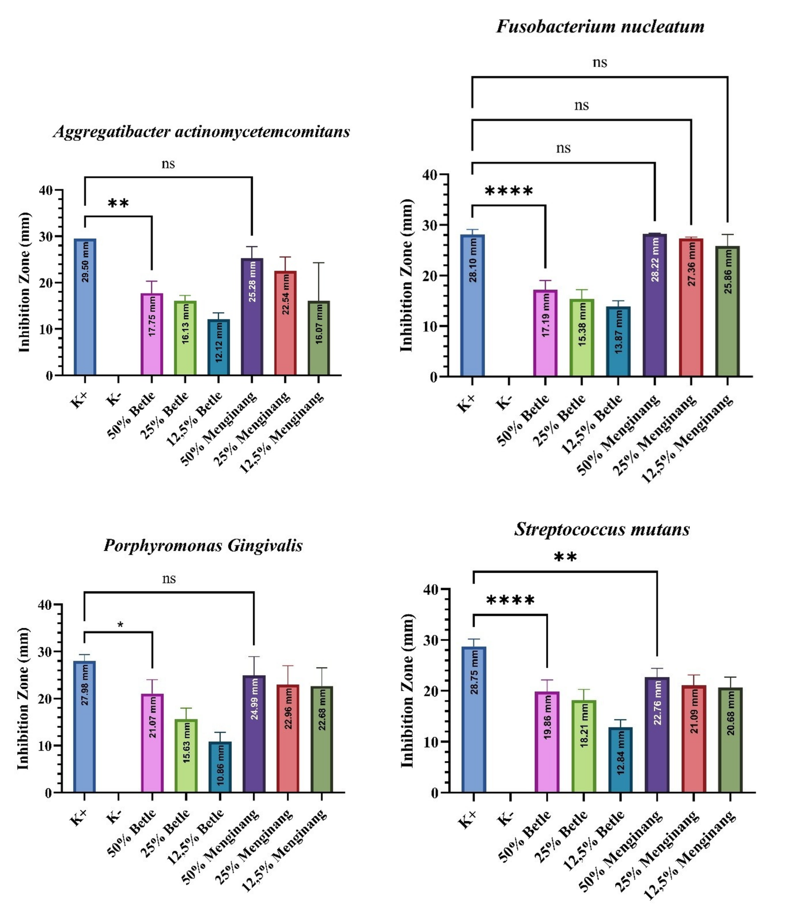
Statistical analysis using Bonferroni’s test (α = 0.05) showed that only the 25% Betle (p = 0.0005) and 12.5% Betle (p < 0.0001) groups had significantly different antibacterial efficacy against P. gingivalis compared to the positive control (K+). Conversely, the 50% Betle (p = 0.0574) and all concentrations of the Menginang group (p > 0.05) showed no significant differences, indicating comparable activity to the positive control.
Bonferroni’s test (α = 0.05) revealed that all Betle concentrations (50%, 25%, and 12.5%) differed significantly from the positive control (K+, p < 0.0001) in inhibitingF. nucleatum. Conversely, all Menginang concentrations showed no significant differences (p > 0.05), indicating antibacterial activity against F. nucleatum that is comparable to K+.
Bonferroni’s test (α = 0.05) showed that all Betle concentrations 50% (p = 0.0039), 25% (p = 0.0011), and 12.5% (p < 0.0001)—differed significantly from the positive control (K+) in inhibiting A. actinomycetemcomitans. In the Menginang group, only the 12.5% concentration showed a significant difference (p = 0.0011), while the 50% and 25% concentrations showed no significant differences (p > 0.05).
Bonferroni’s test (α = 0.05) revealed that all concentrations of both Betle (p < 0.0001) and Menginang (p < 0.01) groups differed significantly from the positive control (K+) in inhibiting S. mutans. This indicates that every evaluated treatment group exhibited a significant difference in antibacterial efficacy compared to K+.
One-Way ANOVA analysis demonstrated statistically significant differences among treatment groups. Post hoc Bonferroni analysis showed that the 50% menginang extract had significantly greater antibacterial activity compared to the lower concentrations and single betel leaf extract groups (p < 0.05).
The effectiveness of plant extracts from the betel-chewing tradition, which is comparable to that of chlorhexidine, can be explained by their similar mechanisms of action. As a cationic biguanide, chlorhexidine strongly binds to negatively charged molecules on the bacterial surface (such as membrane phospholipids and proteins), causing disruption of cell membrane permeability, leakage of protoplasmic contents, inhibition of amino acid and nucleic acid transport enzymes, and ultimately bactericidal cell death at high concentrations (> 0.12%) or bacteriostatic growth inhibition at low doses (28). Chlorhexidine (CHX) is widely recognized as a highly effective antimicrobial agent in dentistry, often referred to as the "gold standard" for chemical plaque control due to its unique property of substantivity the ability to bind to oral surfaces and provide prolonged antimicrobial activity. CHX binds to hydroxyapatite in teeth, dental plaque, and oral tissues, forming a reservoir that releases the antimicrobial agent over time. CHX can also form insoluble compounds with phosphate ions in dental plaque and saliva, further contributing to its prolonged effect (29).
Chlorhexidine exhibits high substantivity by binding to the oral mucosa, dental plaque, and tooth surfaces, followed by gradual release that provides prolonged antibacterial activity against oral pathogens such as S. mutans and P. gingivali (30). This study has several limitations. The antibacterial activity was evaluated only using the disc diffusion method; therefore, the minimum inhibitory concentration (MIC) and minimum bactericidal concentration (MBC) values were not determined. Further studies using MIC and MBC assays are necessary to better evaluate the antibacterial potency and bactericidal properties of the extracts.
Conclusion
This study demonstrated that betel leaf extract (P. betle Linn.) and menginang mixture were able to inhibit the growth of major oral pathogenic bacteria under in vitro conditions. The 50% menginang extract produced the largest inhibition zones among all tested concentrations and extracts approaching that of the positive control 0.2% chlorhexidine. Antibacterial activity is concentration-dependent and may be associated with the presence of multiple bioactive compounds detected in the extracts, including alkaloids, phenolics, flavonoids, tannins, saponins, triterpenoids, and steroids. Thus, betel leaf and menginang extracts demonstrated promising in vitro antibacterial activity against oral pathogenic bacteria against oral pathogenic bacteria in vitro. However, further studies involving toxicity evaluation, formulation optimization, and clinical investigations are necessary before potential therapeutic applications can be considered. Considering its promising antibacterial efficacy, particularly against S. mutans, P. gingivalis, A. actinomycetemcomitans, and F. nucleatum, the menginang extract may be considered for future development as a natural mouthwash or therapeutic oral gel formulation intended to support oral health management.
Declarations
Acknowledgment
The author would like to thank the Faculty of Pharmacy, Muhammad Arsyad Al-Banjari Islamic University of Kalimantan, for providing laboratory facilities as a research site. Thanks are also extended to those who provided technical and administrative support during the implementation of this research. Special gratitude is expressed to apt. Rizki Rahmadi Pratama, M.Farm., for his invaluable guidance and input throughout the research process and the preparation of the results. The author also wishes to acknowledge the Directorate of Learning and Student Affairs, Ministry of Higher Education, Science, and Technology (Kemdiktisaintek), for the support and funding provided through the Student Creativity Program (PKM). Finally, thank you to all members of the PKM team and all parties who cannot be mentioned individually for their direct and indirect support and assistance.
Conflict of Interest
The authors declare no conflict of interest.
Data Availability
Data supporting the findings of this study are available from the corresponding author upon reasonable request.
Ethics Statement
Not applicable.
Funding Information
This work was funded by Student Creativity Program (PKM) under KEMDIKTISAINTEK (Ministry of Higher Education, Science, and Technology) under grant number 2132/B2/DT.01.00/2025.
References
- Purnamasari D, Riyanta AB, Tivani I. Uji aktivitas antibakteri foot sanitizer ekstrak etanol kombinasi lada putih (piperis albi) dan daun teh hijau (camellia sinensis) terhadap bakteri staphylococcus epidermidis. Jkt. 2025;6(1):3775-3782. doi: https://doi.org/10.31004/jkt.v6i1.42967
- Heny Noor Wijayanti. Edukasi Kesehatan Gigi dan Mulut dalam Upaya Meningkatkan Kesehatan Gigi pada Anak Sekolah Dasar. RCS Development. 2023;2(4):153-160. doi: https://doi.org/10.59110/rcsd.201
- Badan Kebijakan Pembangunan Kesehatan, Kementerian Kesehatan Republik Indonesia. Laporan Tematik Survei Kesehatan Indonesia (SKI) 2023: Problematika Kesehatan Gigi dan Mulut di Indonesia. Jakarta: Kementerian Kesehatan Republik Indonesia; 2024.
- Wang B, Deng J, Donati V, Merali N, Frampton AE, Giovannetti E, et al. The Roles and Interactions of Porphyromonas gingivalis and Fusobacterium nucleatum in Oral and Gastrointestinal Carcinogenesis: A Narrative Review. Pathogens. 2024;13(1):93. doi: https://doi.org/10.3390/pathogens13010093
- Rocha CM, Kawamoto D, Martins FH, Bueno MR, Ishikawa KH, Ando-Suguimoto ES, et al. Experimental Inoculation of Aggregatibacter actinomycetemcomitans and Streptococcus gordonii and Its Impact on Alveolar Bone Loss and Oral and Gut Microbiomes. Ijms. 2024;25(15):8090. doi: https://doi.org/10.3390/ijms25158090
- Bonilla M, Martín-Morales N, Gálvez-Rueda R, Raya-Álvarez E, Mesa F. Impact of Protein Citrullination by Periodontal Pathobionts on Oral and Systemic Health: A Systematic Review of Preclinical and Clinical Studies. J Clin Med. 2024;13(22):6831. doi:10.3390/jcm13226831. doi: https://doi.org/10.22541/au.172830784.47641076/v1
- Huang X, Xie M, Lu X, Mei F, Song W, Liu Y, et al. The Roles of Periodontal Bacteria in Atherosclerosis. Ijms. 2023;24(16):12861. doi: https://doi.org/10.3390/ijms241612861
- Gao Z, Chen X, Wang C, Song J, Xu J, Liu X, et al. New strategies and mechanisms for targeting Streptococcus mutans biofilm formation to prevent dental caries: A review. Microbiological Research. 2024;278:127526. doi: https://doi.org/10.1016/j.micres.2023.127526
- Ihsan M. Pembelajaran berbasis Adiwiyata pada siswa kelas VII-2 di Sekolah Menengah Pertama Negeri 9 Parepare [Internet]. Parepare (ID): UPTD SMP Negeri 9 Parepare; 2020 [cited 2026 Jul 9]. Available from: https://doi.org/10.31219/osf.io/96yhr
- Topan Sulaiman Asy’ari, Riska Amelia Safitri, Nur Shofa Fhadila, Nidaul Jannah, Chintana Aurel Hairin, Fauzie Rahman. Menginang Sirih dan Tradisi yang Hampir Hilang pada Masyarakat Banjar serta Perspektifnya dalam Kesehatan Gigi untuk Wujudkan Indonesia Bebas Karies 2030. Health Inf J Penelit. 2023;15(3):e1325. doi: https://doi.org/10.36990/hijp.v15i3.1325
- Eva AFZ, Febriany M, Aslan S, Irawati E, Arifin FA, Fitri NR. Efektivitas Ekstrak Daun Sirih Hijau (Piper betle L.) dalam Menghambat Pertumbuhan Bakteri Streptococcus mutans. Smj. 2023;5(01):32-38. doi: https://doi.org/10.33096/smj.v5i01.95
- Afifah Rukmini. Skrining fitokimia familia piperaceae. JB&P. 2020;7(1):28-32. doi: https://doi.org/10.29407/jbp.v7i1.14805
- Handoyo DLY. The Influence Of Maseration Time (Immeration) On The Vocity Of Birthleaf Extract (Piper Betle). Tinctura. 2020;2(1):34-41. doi: https://doi.org/10.35316/tinctura.v2i1.1546
- Hasanuddin ARP, Salnus S. Uji bioaktivitas minyak cengkeh (Syzygium aromaticum) terhadap pertumbuhan bakteri Streptococcus mutans penyebab karier gigi. Bioma J Biol Makassar. 2020;5(2):241–250. doi:10.20956/bioma.v5i2.11490
- Arifin SHAG, Firdhausi NF, Hidayati I, Faizah H. Phytochemical Screening and Antimicrobial Activity of Combination of Piper betle and Moringa oleifera Extracts. Ijpst. 2025;12(2):184-191. doi: https://doi.org/10.24198/ijpst.v12i2.45288
- Mulyaningsih S, Ria Indah Pratami, Ichwan Ridwan Rais. Optimization of Water-Ethanol Solvent Composition of Red Betel Leaf Extraction (Piper crocatum Ruiz & Pav) Using Simplex Latice Design Method on Antioxidant Activity. Jfm. 2025;8(1):311-319. doi: https://doi.org/10.35451/5d1c7339
- Pasaribu RKU, Iqbal M, Rahayu ID, Afriyani, Triyandi R. Pengaruh Pemilihan Pelarut Terhadap Rendemen Ekstrak Daun Teh Hijau(Camellia sinensis) Menggunakan Metode Ekstraksi Maserasi. SnsMed. 2025;3(5):275-279. doi: https://doi.org/10.63004/snsmed.v3i5.734
- Sun S, Yu Y, Jo Y, Han JH, Xue Y, Cho M, et al. Impact of extraction techniques on phytochemical composition and bioactivity of natural product mixtures. Front. Pharmacol. 2025;16:1615338. doi: https://doi.org/10.3389/fphar.2025.1615338
- Jam N, Hajimohammadi R, Gharbani P, Mehrizad A. Evaluation of Antibacterial Activity of Aqueous, Ethanolic and Methanolic Extracts of Areca Nut Fruit on Selected Bacteria. BioMed Research International. 2021;2021(1):6663399. doi: https://doi.org/10.1155/2021/6663399
- Nayeem J, Dey SK, Dey P, Himel IA, Khatoon H. A comprehensive dataset on antimicrobial activity of indigenous Oscillatoria spp. against common bacteria causing diseases in fish and shellfish. Data in Brief. 2023;50:109563. doi: https://doi.org/10.1016/j.dib.2023.109563
- Okonogi S, Phumat P, Khongkhunthian S. Enhancement of aqueous solubility and antibiofilm activity of 4-allylpyrocatechol by polymeric micelles. Bioprocess Biosyst Eng. 2021;44(6):1289-1300. doi: https://doi.org/10.1007/s00449-020-02501-7
- Ratchasong K, Saengsawang P, Yusakul G, Makkliang F, Lakhanapuram HK, Wintachai P, et al. Bactericidal Activities of Nanoemulsion Containing Piper betle L. Leaf and Hydroxychavicol Against Avian Pathogenic Escherichia coli and Modelling Simulation of Hydroxychavicol Against Bacterial Cell Division Proteins. Antibiotics. 2025;14(8):788. doi: https://doi.org/10.3390/antibiotics14080788
- Nayaka NMDMW, Sasadara MMV, Sanjaya DA, Yuda PESK, Dewi NLKAA, Cahyaningsih E, et al. Piper betle (L): Recent Review of Antibacterial and Antifungal Properties, Safety Profiles, and Commercial Applications. Molecules. 2021;26(8):2321. doi: https://doi.org/10.3390/molecules26082321
- Krongkeha W, Pitaktim S. Growth inhibition of Escherichia coli and Pseudomonas aeruginosa strains by Piper betle Linn. extracts. J Curr Sci Technol. 2022;12(1):141–150. doi:10.14456/jcst.2022.13
- Amrillah N, Triyandi R, Iqbal M, Pardilawati CY. Literature study: antibacterial activity of betel leaf extract (Piper betle L.) against Staphylococcus aureus. Medula. 2023;13(4):578–582. doi:10.53089/medula.v13i4.754
- Proskura A, Muradova M, Poirier N, Ilina V, Chaloyard J, Boichot V, et al. The Ability of Basic and Glycosylated Proline-Rich Proteins to Protect Oral Enzymes from Polyphenol Inhibition. J. Agric. Food Chem. 2025;73(33):21000-21008. doi: https://doi.org/10.1021/acs.jafc.5c00821
- Seukep AJ, Ojong OCG, Mbuntcha HG, Matieta VY, Zeuko’o EM, Kouam AF, et al. In Vitro Antibacterial Potential of Herbal Beverage Extracts From Cinnamon, Clove, and Thyme and Their Interactive Antimicrobial Profile With Selected Antibiotics Against Drug‐Resistant Clinical Pathogens. Journal of Tropical Medicine. 2025;2025(1):9916282 (17 p). doi: https://doi.org/10.1155/jotm/9916282
- Widodo ADS, Setyaningsih S, Astuti P, Meilawaty Z, Suci Dharmayanti AW. Uji efektivitas antibakteri daun Biduri (Calotropis gigantea) terhadap pertumbuhan Streptococcus mutans: studi eksperimental laboratoris. J Ked Gi Unpad. 2025;36(3):312-320. doi: https://doi.org/10.24198/jkg.v36i3.56148
- Thangavelu A, Kaspar S, Kathirvelu R, Srinivasan B, Srinivasan S, Sundram R. Chlorhexidine: An elixir for periodontics. J Pharm Bioall Sci. 2020;12(5):57. doi: https://doi.org/10.4103/jpbs.jpbs_162_20
- Poppolo Deus F, Ouanounou A. Chlorhexidine in Dentistry: Pharmacology, Uses, and Adverse Effects. International Dental Journal. 2022;72(3):269-277. doi: https://doi.org/10.1016/j.identj.2022.01.005