RESEARCH ARTICLE
In Vitro Evaluation of the Antifungal Activity of Bioactive Extract of Argemone mexicana against Fusarium oxysporum Associated with Ginger Rhizome Rot
Academic Editor: Samir Chtita
Sciences of Phytochemistry|Vol. 5, Issue 2, pp. 359-367 (2026)
CC BY 4.0-2026 Authors

Received
Jul 23, 2026Revised
Aug 26, 2026Accepted
Oct 10, 2026Published
Nov 12, 2026
Abstract
Plant bioactives are emerging as environment-friendly antifungal agents and as safer alternative to chemical fungicides. Fusarium oxysporum is one of major phytopathogens infecting ginger rhizomes. This fungal species predominates in soil and, under moist conditions, damages seeds or rhizomes. Available data indicated that Argemone mexicana exhibits antifungal properties; however, this plant has not been systematically studied against F. oxysporum, the causative agent of rhizome rot disease of ginger. The present study aimed to evaluate the antifungal activity of the bioactive extract of A. mexicana against F. oxysporum. Fungal species from infected ginger were systematically characterized by microscopic and molecular techniques, including ITS-based molecular identification and BLAST analysis. The plantwas collected from the Melghat forest. Preparation of leaf extract of plants followed by preliminary phytochemical characterization where TLC showed distinct bands with Rf values of 0.12, 0.16, 0.21, and 0.79, and HPLC showed a prominent peak at retention time 7.863, supporting the presence of alkaloids in the ethanolic leaf extract. Further, bioactive ethanolic leaf extract of A. mexicana at a concentration of ≥3 mg/mL inhibited mycelial growth even after seven days of incubation, while no visible mycelial growth was observed at 1–2 mg/mL up to day 4 of incubation, after which limited growth appeared. The study indicated that A. mexicana leaf extract exhibited a pronounced effect against F. oxysporum proliferation, which can be an alternative in agricultural practices.
Keywords:
Introduction
A faster increase in the human population has led to excessive use of synthetic fertilizers and pesticides to meet global food demand. Productivity and yield of crops are critically affected by numerous insects and pests, encouraging agriculturists to adopt various pest control strategies. Frequent and casual use of chemical pesticides has become general in modern agricultural applications. The majority of pesticidal compounds are made up of hazardous chemicals, and inappropriate applications of these compounds have been associated with negative impacts on human health, and on fauna and terrestrial and aquatic environments (1, 2). Fungal infections account for several plant diseases and for a remarkable loss in production yield of crops up to 14% every year, which is significantly responsible in the context of agricultural sustainability and food security around the globe (3). Among the fungal isolates, Fusarium oxysporum was selected because it is a widespread and economically important plant pathogen. It causes Fusarium wilt in many crops, leading to poor plant growth and significant yield losses. Its broad host range and ability to persist in soil make it difficult to control. Therefore, F. oxysporum was considered a relevant target for evaluating potential antifungal agents and developing more sustainable disease management approaches. F. oxysporum is particularly known as causative agent responsible for vascular wilt and root rot disease in more than 100 agriculturally significant crops (4). The fungicides, made up of harsh chemicals, are commonly applied directly to the soil by means of seed treatment before sowing for the inhibition of fungal pathogens. However, routine usage of synthetic fungicides leads to the development of resistance in fungal pathogens, accumulation of toxic residues, environmental persistence and health-related risks (5). Additionally, the economic impact and lack of harmony with organic farming practices reduce their sustainability. Thus, naturally derived products, especially bioactive compounds of plant origin are being investigated as eco-friendly and safer alternatives for the management of fungal pathogens (6). Ethanolic leaf extracts of Tridax procumbens and Xanthium strumarium exhibited antifungal activity, suggesting the potential of plant-based formulations as a sustainable fungicide (7, 8).
Among the plant-derived sources explored for antifungal activity, Argemone mexicana L. has attracted attention as a promising candidate due to its rich and diverse phytochemical composition. Belonging to the family Papaveraceae, Argemone mexicana is an annual herb native to Mexico and tropical regions of America, but it is now widely distributed across tropical and subtropical areas, including India, where it commonly grows as a weed. Despite its widespread occurrence, the plant contains a variety of bioactive compounds, including alkaloids, phenolic compounds, and other secondary metabolites that have been linked with antimicrobial activity. Earlier studies have also reported the antifungal potential of its phytochemical constituents against several phytopathogenic fungi. Taken together, these findings suggest that Argemone mexicana may serve as a useful natural source of antifungal compounds and provide a reasonable scientific basis for evaluating its activity against F. oxysporum, a major pathogen associated with ginger rhizome rot. The plant has been used in traditional medicines since ancient times for skin disease management (9), inflammatory disorders, jaundice and various infectious diseases. Various parts of Argemone mexicana possess wide therapeutic properties like anti-malarial (10, 11) as well as wound-healing (12). Modern scientific evidence highlights Argemone mexicana as a potent and rich source of bioactive components with pharmacological, broad-spectrum antibacterial importance (13, 14).
The mixture of quaternary alkaloids from Argemone mexicana exhibited efficient antifungal activity against numerous phytopathogenic fungi with significant inhibition of spore germination (15). The plant formulations of Argemone mexicana demonstrated remarkable actions as botanical fungicides (16). The management of fungal pathogens illustrates a major challenge for farmers.
Previous studies have shown that Argemone mexicana and its phytochemical constituents possess promising antimicrobial and antifungal properties. The plant is reported to contain numerous secondary metabolites, including alkaloids, phenolics, flavonoids, terpenoids and many more, with several biological activities (17, 18). Alkaloids are major compounds in Argemone mexicana; sanguinarine, chelerythrine, berberine, protopine and allocryptopine are a group of benzylisoquinoline alkaloidsare known to possess antimicrobial and other biological actions (19). These phytochemical attributes provide a solid scientific platform for investigation of the efficacy of Argemone mexicana against phytopathogenic fungi. Therefore, the present study was designed to evaluate whether the bioactive constituents of Argemone mexicana could inhibit F. oxysporum and provide a potential plant-based alternative for fungal disease management in ginger. However, its activity against F. oxysporum specifically associated with ginger rhizome rot has received comparatively little attention. Therefore, the present study focused on isolating and identifying the fungal pathogen from diseased ginger rhizomes through morphological characterization and ITS-based molecular analysis. The identified F. oxysporum was then used to evaluate the in vitro antifungal activity of an ethanolic leaf extract of Argemone mexicana. Along with this, the phytochemical profile of the extract was investigated to gain a better understanding of its bioactive constituents and their possible contribution to antifungal activity. Overall, the study aimed to explore the potential of Argemone mexicana as a natural, plant-derived option for managing fungal pathogens affecting ginger.
Materials & Methods
Isolation of Fungus From Infected Ginger Crop
The ginger (Zingiber officinale Rosc.) with symptoms of disease was identified and collected from farmland at 20.0576º N and 76.5662º E. Tissues with infection were found to be soft due to pathogen activity. The samples were collected in sterile sampling bags and transported to the laboratory. In the laboratory, the collected samples were carefully washed and treated with NaOCl for the removal of soil, dirt and adhesive particles. Infected tissues were inoculated onto potato dextrose agar (PDA) aseptically and incubated at 27 °C for four days for complete growth of fungi. Thereafter, they were sub-cultured onto fresh PDA media to obtain a pure fungal isolate, which was used for further experimental analysis.
Characterization of Isolated Fungus
Morphological Characterization of Isolated Fungus
Staining procedures were performed using lactophenol cotton blue stain to determine the micromorphological characters of the fungal isolate. Structure of hyphae, conidia and septation patterns were considered for identification
Molecular Characterization Using Its Region Sequencing
Fungal growth was undertaken for extraction of genomic DNA; after extraction, partial amplification of the extracted region was performed using universal primers ITS1 and ITS4. PCR amplification resulted in a distinct single band on 1.3% agarose gel. An amplified fragment was extracted, purified and put through bidirectional sequencing using forward and reverse primers. The acquired ITS region sequence was compared to sequences available in the NCBI GenBank database using the Basic Local Alignment Search Tool (BLAST) to determine the closely related taxa on the basis of similarity of sequence and quality of alignment. Using ClustalW, multiple sequence alignment was performed, and a distance matrix was generated. Phylogenetic tree construction and analysis were done using MEGA 12 for determination of the taxonomic position of the isolate (20).
Herbarium Preparation and Deposition
A.mexicana with flower, fruit and vegetative structure was collected and mounted over blotting paper. For proper drying and pressing, the mounted specimen was kept in wooden blocks. The specimen was mounted on a standard herbarium sheet with proper labels including botanical name, locality, collection dates, etc. (21). The herbarium prepared by standard procedures was then deposited at the Botanical Survey of India (BSI), Pune.
Collection of Plant Leaves
The leaves were collected from healthy plants from the Melghat forest of Amravati, at a latitude of 21.378º N and longitude of 77.348º E, transported to the laboratory, followed by washing the leaves under running tap water to remove dust and soil particles from the surface of the leaves. After washing, the leaves were shade-dried; the dried leaves were ground to form fine powder, the powder was strained through a mesh and stored in airtight containers for further processing.
Preparation of Crude Extract
The leaf powder was extracted using 95% ethanol by the cold maceration method. Approximately 50 g of leaf powder was immersed in ethanol and kept on a rotary shaker for 72 h. The resulting mixture was filtered through Whatman filter paper, and the filtrate was collected and concentrated by drying in a hot air oven at temperatures below 40 °C. The dried extract was powdered to form uniform fine particles and stored at 4 °C until further use in biological assays.
Identification of Phytochemicals: Alkaloids
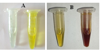
Phytochemical analysis of the extract was carried out for assessment of the persistence of phytochemical compounds like alkaloids. Wagner’s test was conducted for the identification, where a potassium iodide (2 g) and iodine (1.27 g) solution in 100 mL distilled water was prepared. A few drops of this solution were added to the filtrate; formation of a brown-colored precipitate indicated the presence of alkaloids. (22).
Hager’s test was performed as 0.02 gm of leaf extract of Argemone mexicana was weighed in 3 different test tubes. 10 mL of methanol was added to each test tube, and the test tubes were placed in a vortex mixer to dissolve the extracts. The extract solutions were then filtered. 2 mL of filtrate was taken and mixed with 4 drops of 2% H2SO4. To 1 mL of this mixture, 6 drops of Hager´s reagent were added. Yellow (turbid) color indicates the presence of alkaloids (22).
Thin Layer Chromatography (TLC)
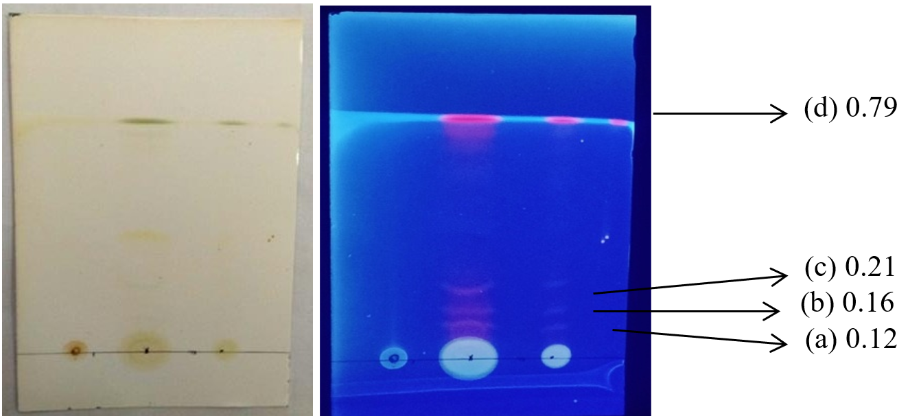
By reconstituting the 1 mg of ethanolic extract in 2 mL of methanol, the TLC was performed. A precoated Silica gel 60 F254 sheet (10x10 cm) was used for TLC. The mobile phase of benzene: ethanol (3:1.5) mL was used for each spot. After the solvent reached the front, the plate was dried for a few seconds, then visualized under a UV-transilluminator for determination of the resultant band at 365 nm (23, 24, 25). The TLC analysis was performed using the previously reported solvent system containing benzene. Considering the recognized safety hazards associated with benzene, safer validated solvent alternatives should be considered for future phytochemical analyses.
High Performance Liquid Chromatography (HPLC)
HPLC analysis was carried out to confirm the presence of phytoconstituents in the extracts. Around 10 mg plant extract was dissolved in 100 mL of methanol to get a concentration of 100 μg/mL. Then samples were passed through nylon syringe filter. HPLC was carried out on Waters 1525 Binary HPLC pump and Waters 2998 photodiode array detector. The sample was separated by chromatographic separator Water C-18 column. Breeze-2 HPLC software was used for integration and calibration. The mobile phase of acetonitrile: water in a ratio of 10:90 was used (26, 27, 28).
Evaluation of Antifungal Activity by the Poisoned Food Technique
Antifungal activity of the ethanolic extract of Argemone mexicana was evaluated using the qualitative poisoned-food technique. For this, different concentrations of crude ethanolic extract were prepared in 20% ethanol to check the antifungal activity of the plant extract. The extract was prepared at concentrations of 10, 20, 30, 40, 50 and 60 mg/mL. For the poisoned food assay, 1 mL of each concentration was aseptically added to 9 mL of molten PDA, giving final concentrations 1, 2, 3, 4, 5 and 6 mg/mL in the assay medium; likewise, 1 mL of 20% ethanol and 1 mL of 10 mg/mL copper oxychloride added to molten PDA; and considered as control and standard for the assay respectively (29). After solidification at room temperature, a 5 mm diameter mycelial disc was cut from the actively growing margin of a 7-day-old F. oxysporum culture and placed on the poisoned PDA plate. Incubation was done at 27 °C for 5-7 days, and fungal inhibition was observed and recorded (30).
Results
Isolation and Morphological Characterization of Fungi
Ginger rhizomes exhibiting characteristic disease symptoms were collected and processed for fungal isolation. Samples were rinsed under sterile conditions to remove surface contaminants. Small sections from infected tissues were aseptically placed on potato dextrose agar (PDA) and incubated under controlled conditions. Fungal growth became evident within 72 h, indicating the presence of viable fungal pathogens (Figure 1).
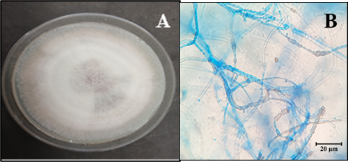
The isolate, which formed concentric brownish-white mycelial growth, was obtained. Microscopic characteristics were examined by staining with lactophenol cotton blue. The observations revealed conidial structure and hyphal morphologies of Fusarium species. Based on cultural and microscopic properties, the isolate was identified as a species of Fusarium; later, it was supported by molecular characterization.
Molecular Characterization of Fungal Isolates by Its Region Sequencing
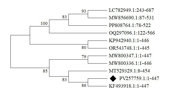
PCR amplification of ITS region using universal primers resulted in a single, distinct band of expected size with high-quality DNA. Purification and sequencing of the amplified product were done, followed by BLAST using the NCBI database to identify the isolate. As per the Phylogenetic analysis, the obtained sequence showed high-level similarity with the reference sequence of F. oxysporum, revealing the taxonomic position of the isolate (Figure 2). The close matches showed high query coverage, strong alignment scores, as well as minimal nucleotide variations. To ease data availability and reproducibility, the authenticated ITS sequence was deposited in the NCBI GenBank database under the accession number PV257759.1, providing a permanent molecular reference for this isolate.
Herbarium Preparation and Deposition
According to standard procedures for herbarium preparation, the herbarium specimen of Argemone mexicana was prepared. The specimen was deposited as specimen number 02 with reference number BSI/WRC/Tech./2025/JVD-196, for proper documentation, reproducibility, and reference for further studies.
Collection, Extraction of Leaves of Argemone mexicana, and Identification of Bioactive Phytocompound (Alkaloid)
Fresh leaves from healthy Argemone mexicana plants were collected from the Melghat forest of Amravati, Maharashtra, India, with prior permission from the Maharashtra State Biodiversity Board (MSBB), Nagpur. Plant material with no traces of insect appearance was selected to avoid microbial contaminants and quality of bioactives. The collected leaves were washed with clean and running tap water for removal of dust debris, followed by shade-drying at normal temperature. After complete drying, leaves were ground into homogeneous powder and strained through a mesh sieve. Powdered plant leaves were used for extraction using ethanol, in a 1:5 ratio, as it has the potential to dissolve a wide range of polar as well as partially non-polar phytoconstituents. Crude ethanolic extract was the outcome of the extraction process; the extract was then concentrated, dried and finally used for antifungal activity assessment.
TLC and HPLC Characterization of Extracted Compound
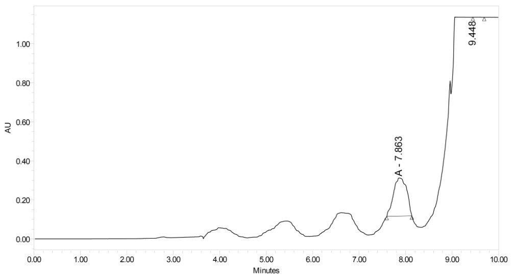
From the TLC and HPLC analysis of the extract, the Rf values of the bands observed by TLC (Figure 4) and the highest peak at a retention time of 7.863 min in HPLC (Figure 5), where the X-axis represents time and the Y-axis represents absorbance in absorbance units (AU), were recorded. The results showed resemblance to previous analyses, and thus the presence of alkaloids in the plant extract of Argemone mexicana was determined.
Evaluation of Antifungal Activity by the Poisoned Food Technique
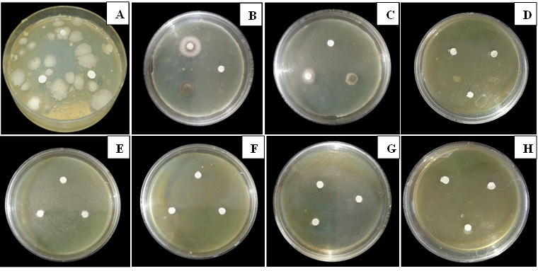
Antifungal activity of bioactive ethanolic extract from Argemone mexicana was evaluated using the poisoned food technique by means of examining mycelial growth of the fungus at different concentrations of the extract at different intervals of time. Copper oxychloride, a commercially available fungicide, was used as a positive control (Standard). Ethanolic extract was found to show a concentration-dependent inhibitory effect against fungal growth. Initially, no visible mycelial growth was observed even at lower concentrations (1-2 mg/mL), which is an indication of the suppressive influence of the extract on fungal growth. After four days of incubation, limited mycelial growth appeared at these initial concentrations.
Higher concentrations of extract showed sustained and remarkable antifungal potential. Extract concentrations above 3 mg/mL and the positive control showed complete inhibition of mycelial growth and development even after seven days of incubation, demonstrating strong and consistent antifungal activity.
In the poisoned food assay, the test extract was incorporated into the medium, i.e. PDA itself, before inoculation, and the mycelial discs were placed on the medium. The three spots on each plate represent separate inoculation points and should not be interpreted as diffusion discs. Substantially, the present findings demonstrate the dose-dependent antifungal potential of Argemone mexicana, with higher concentrations showing efficient and sustained inhibition of fungal mycelial growth (Figure 6).
Discussion
The isolated fungus from the infected ginger was identified as F. oxysporum based on the cultural characteristics, mycelial growth pattern and microscopic observations. In microscopy, when stained with lactophenol cotton blue, it preliminarily describes the morphological characteristics of Fusarium sp. (7, 8, 31). In the present study, the fungal isolate was identified as F. oxysporum based on ITS sequence analysis using the ITS1 and ITS4 primers, and the sequence was deposited in GenBank under accession number PV257759.1. Although the ITS region is widely used for fungal identification, its limited discriminatory power within the F. oxysporum species complex may restrict reliable species-level identification. Protein-coding markers, particularly the translation elongation factor 1-alpha (TEF-1α) gene, generally provide greater phylogenetic resolution. Therefore, the ITS-based identification reported in this study should be interpreted with caution, and future multilocus sequence analysis using TEF-1α and additional informative markers could provide stronger confirmation of the taxonomic identity of the isolate.
Other fungal pathogens that infect ginger have been characterized as Fusarium and were found to be inhibited by extracts of T. procumbens (7). Root-infecting fungal pathogens were inhibited using extracts of Argemone mexicana and fungal species, including Fusarium solani and Rhizoctonia solani (32). Argemone mexicana extracts were found to be effective against some species of Candida, including Candida albicans, Candida stellatoidea, Candida torulopsis, etc., which indicates potential of Argemone mexicana against human pathogens as well (33).
The antifungal testing described fungistatic as well as significant fungicidal potential of Argemone mexicana at different concentrations of ethanolic extracts against phytopathogenic F. oxysporum. The extract contained numerous active components with efficient fungicidal potential. As F. oxysporum is one of the major soil-borne fungal pathogens and causative agent of numerous infections in ginger leading to Fusarium rot as well as Fusarium wilt disease; observed fungicidal activity of extracts from Argemone mexicana might be a promising sustainable alternative for fungal pest management. Aqueous and methanolic extracts of Argemone mexicana exhibited inhibitory effects against Mucor indicus, Aspergillus flavus, Aspergillus niger and Penicillium notatum (34).
Antifungal efficacy of Argemone mexicana observed in this study shows consistency with earlier findings demonstrating broad-spectrum antimicrobial activities. As per the reports of (16), Argemone mexicana extracts reduced mycelial growth and spore formation of F. solani. The findings of (7) also revealed the potential of T. procumbens plant extracts against Fusarium sp.isolated from infected ginger and highlight the antifungal potential of plant-based green fungicides. The fungal growth-inhibitory potential of plant extracts may be due to the presence of alkaloids, flavonoids, phenolics and terpenoids in the plant extracts.
The concurrence of present findings with the earlier studies supports the antifungal potential of bioactive phytochemicals from Argemone mexicana. Currently, plant-derived pesticides are getting wide attention as they are natural, safe and sustainable alternatives to synthetic pest control agents. In this context, the activity of plant formulations of Argemone mexicana against F. oxysporum attributes its potential as a natural antifungal.
Conclusion
The present study demonstrated that the ethanolic leaf extract of Argemone mexicana showed remarkable antifungal activity against the Fusarium sp. isolated from soft rot of ginger. The observed inhibition, compared with copper oxychloride, revealed that Argemone mexicana could be a promising source for plant-based antifungal compounds. However, the findings of the present study are based on in vitro analysis, and the bioactive compounds responsible for the antifungal effect were not characterized completely. Further studies should therefore focus on identification of the major bioactive compounds, determination of the mode of action and evaluation of the effectiveness of the bioactives under greenhouse conditions as well as in fields. Overall, the findings of the study provide a useful preliminary basis for exploring Argemone mexicana as a promising and efficient component of safer and sustainable approaches for the management of fungal diseases of ginger.
Declarations
Acknowledgment
The authors acknowledge Honorable Vice-Chancellor, Sant Gadge Baba Amravati University, Amravati, for providing infrastructure facilities during the research. Mahatma Jyotiba Phule Research and Training Institute (MAHAJYOTI), Nagpur, is acknowledged for providing research fellowship to first author. No AI was used in the writing or during the study.
Conflict of Interest
The authors declare no conflict of interest.
Data Availability
All data generated or analyzed during this study are included in this published article and its supplementary information files.
Ethics Statement
Not applicable.
Funding Information
This work received no external funding.
References
- Sabarwal A, Kumar K, Singh RP. Hazardous effects of chemical pesticides on human health–Cancer and other associated disorders. Environmental Toxicology and Pharmacology. 2018;63:103-114. doi: https://doi.org/10.1016/j.etap.2018.08.018
- Zubrod JP, Bundschuh M, Arts G, Brühl CA, Imfeld G, Knäbel A, et al. Fungicides: An Overlooked Pesticide Class?. Environmental Science & Technology. 2019;53(7):3347-3365. doi: https://doi.org/10.1021/acs.est.8b04392
- Anand G, Rajeshkumar KC. Challenges and Threats Posed by Plant Pathogenic Fungi on Agricultural Productivity and Economy. Singapore: Springer Nature Singapore; 2022. doi: https://doi.org/10.1007/978-981-16-8877-5_23
- Li PL, Shi YX, Guo MY, Xie XW, Chai AL, Li BJ. Fusarium wilt of cauliflower caused by Fusarium equiseti in China. Canadian Journal of Plant Pathology. 2017;39(1):77-82. doi: https://doi.org/10.1080/07060661.2017.1301998
- van Rhijn N, Rhodes J. Evolution of antifungal resistance in the environment. Nat Microbiol. 2025;10(8):1804-1815. doi: https://doi.org/10.1038/s41564-025-02055-y
- Jiménez-Reyes MF, Carrasco H, Olea AF, Silva-Moreno E. Natural compounds: a sustainable alternative to the phytopathogens control. J. Chil. Chem. Soc. 2019;64(2):4459-4465. doi: https://doi.org/10.4067/s0717-97072019000204459
- Awachar S, Patil BS, Charpe P, Kamble KD. Sustainable management of Fusarium-induced rhizome rot in ginger using bioactive extracts of Tridax procumbens. Asian Jr. of Microbiol. Biotech. Env. Sc. 2026;28(1-2):57-62. doi: https://doi.org/10.53550/ajmbes.2026.v28.i01-02.008
- Awachar S, Patil B, Kamble K. In vitro antifungal activity of Xanthium strumarium derived xanthinin against Ginger-infecting Fusarium oxysporum. Res. J. Biotech. 2026;6(21):29. doi: https://doi.org/10.25303/216rjbt029034
- Rageh RS, Algfri SK, Naser GA, Shuaib AB. Evaluation of pharmacognostic, phytochemical, and antioxidant properties of Argemone mexicana linn leaves. J. Med. Plants Stud. 2024;12(6):36-43. doi: https://doi.org/10.22271/plants.2024.v12.i6a.1765
- Willcox ML, Graz B, Falquet J, Sidibé O, Forster M, Diallo D. Argemone mexicana decoction for the treatment of uncomplicated falciparum malaria. Transactions of the Royal Society of Tropical Medicine and Hygiene. 2007;101(12):1190-1198. doi: https://doi.org/10.1016/j.trstmh.2007.05.017
- Simoes-Pires C, Hostettmann K, Haouala A, Cuendet M, Falquet J, Graz B, et al. Reverse pharmacology for developing an anti-malarial phytomedicine. The example of Argemone mexicana. International Journal for Parasitology: Drugs and Drug Resistance. 2014;4(3):338-346. doi: https://doi.org/10.1016/j.ijpddr.2014.07.001
- Mengie Ayele T, Chekol Abebe E, Tilahun Muche Z, Mekonnen Agidew M, Shumet Yimer Y, Tesfaw Addis G, et al. Evaluation of In Vivo Wound-Healing and Anti-Inflammatory Activities of Solvent Fractions of Fruits of Argemone mexicana L. (Papaveraceae). Evidence-Based Complementary and Alternative Medicine. 2022;2022:1-17. doi: https://doi.org/10.1155/2022/6154560
- Priya CL, Rao KV. Ethanobotanical and current ethanopharmacological aspects of Argemone mexicana Linn: an overview. International Journal of Pharmaceutical Sciences and Research. 2012;3(7):2143. url: https://www.cabidigitallibrary.org/doi/full/10.5555/20123285435
- More NV, Kharat KR, Kharat AS. Berberine from Argemone mexicana L exhibits a broadspectrum antibacterial activity. Acta Biochim Pol. 2017;64(4):653-660. doi: https://doi.org/10.18388/abp.2017_1621
- Singh S, Singh A, Jaiswal J, Singh TD, Singh VP, Pandey VB, et al. Antifungal activity of the mixture of quaternary alkaloids isolated from Argemone mexicana against some phytopathogenic fungi. Archives Of Phytopathology And Plant Protection. 2010;43(8):769-774. doi: https://doi.org/10.1080/03235400802176159
- Patil BJ, Kamble SK. Inhibitory effect of some plant extracts against urediospore germination of Uromyces ciceris–arietini (Grognot) Jacz. & Boyer. International Journal of Research in Plant Science. 2014;4(3):60-2.
- Sharanappa R, and Vidyasagar, GM. Documentation of traditional knowledge on medicinal plants used in treating candidiasis in Hyderabad Karnataka region, India. World Journal of Pharmacy and Pharmaceutical Sciences. (2014); 3 (7): 739-751. url: https://www.cabidigitallibrary.org/doi/full/10.5555/20143284054
- Patocka J, Ovando-Martinez M, Navratilova Z, Oleksak P, Ogwu MC, Nepovimova E, et al. Argemone mexicana L. (Papaveraceae): chemistry, pharmacology, and toxicology. Phytochem Rev. 2024;23(4):1167-1182. doi: https://doi.org/10.1007/s11101-024-09918-y
- Brahmachari G, Gorai D, Roy R. Argemone mexicana: Chemical and pharmacological aspects. Revista Brasileira de Farmacognosia. 2013;23(3):559-575. doi: https://doi.org/10.1590/s0102-695x2013005000021
- Kumar S, Stecher G, Suleski M, Sanderford M, Sharma S, Tamura K. MEGA12: Molecular Evolutionary Genetic Analysis version 12 for adaptive and green computing. Molecular Biology and Evolution. 2024; 41 (12):1-9. doi: https://doi.org/10.1101/2024.12.10.627672
- Bhandari JB, Gurung C. Instrumentations Manual in Biology. New Delhi: Narosa Publishing House; 2020. p. 114.
- Singh V, Kumar R. Study of phytochemical analysis and antioxidant activity of Allium sativum of Bundelkhand region. International Journal of Life-Sciences Scientific Research. 2017 Nov;3(6):1451-8. doi: https://doi.org/10.21276/ijlssr.2017.3.6.4.
- Alam SD, Beg MA, Bagadi M, Locatelli M, ALOthman ZA, Mustaqeem M, et al. Facile extraction of berberine from different plants, separation, and identification by thin‐layer chromatography, high‐performance liquid chromatography, and biological evaluation against Leishmaniosis. J of Separation Science. 2023;46(21):2300582. doi: https://doi.org/10.1002/jssc.202300582
- Goswami AK, Gogoi N, Shakya A, Sharma HK. Development and Validation of High-Performance Thin-layer Chromatographic Method for Quantification of Berberine in Rhizomes of Coptis teeta Wall, an Endangered Species Collected from Arunachal Pradesh, India. Journal of Chromatographic Science. 2019;57(5):411-417. doi: https://doi.org/10.1093/chromsci/bmz009
- Monforte-González M, Guízar-González C, Rubio-Piña J, Carrillo-Pech M, Vázquez-Flota F. Berberine and Sanguinarine quantitation inArgemone mexicanaL. (Papaveraceae) tissues by TLC-in situfluorography. Journal of Planar Chromatography – Modern TLC. 2012;25(4):358-360. doi: https://doi.org/10.1556/jpc.25.2012.4.14
- Satija S, Malik S, Garg M. Development of a new, rapid, and sensitive validated high-performance thin-layer chromatographic method for the estimation of berberine inTinospora cordifolia. JPC - Journal of Planar Chromatography - Modern TLC. 2016;29(3):209-215. doi: https://doi.org/10.1556/1006.2016.29.3.7
- Srinivasan G, Unnikrishnan K, Rema Shree A, Balachandran I. HPLC estimation of berberine in <i> Tinospora cordifolia</i> and <i> Tinospora sinensis</i>. Indian J Pharm Sci. 2008;70(1):96. doi: https://doi.org/10.4103/0250-474x.40341
- Wang K, Chai L, Feng X, Liu Z, Liu H, Ding L, et al. Metabolites identification of berberine in rats using ultra-high performance liquid chromatography/quadrupole time-of-flight mass spectrometry. Journal of Pharmaceutical and Biomedical Analysis. 2017;139:73-86. doi: https://doi.org/10.1016/j.jpba.2017.02.038
- Singh DK, Singh R. Antifungal activity of ethanolic extracts of Eupatorium adenophorum leaves. Indian Journal in Pharmacy and Biotechnology. 2013;1(4):562-4.
- Devkota A, Das RK. Antibacterial Activities of X. strumarium L. J. Nat. Hist. Mus. 2018;29:70-77. doi: https://doi.org/10.3126/jnhm.v29i0.19039
- Harsha K, Nair AR, Tejaswini G. Identification of F. oxysporum as causative agent of rhizome rot disease in seed ginger rhizomes in Wayanad, Kerala. Indian Phytopathology. 2021;74(1):253-256. doi: https://doi.org/10.1007/s42360-020-00315-x
- Siddiqui IA, Shaukat SS, Khan GH, Zaki MJ. Evaluation of Argemone mexicana for Control of Root‐Infecting Fungi in Tomato. Journal of Phytopathology. 2002;150(6):321-329. doi: https://doi.org/10.1046/j.1439-0434.2002.00762.x
- Osho A, Adetunji T. Antimicrobial activity of the essential oil of Argemone mexicana Linn. Journal of Medicinal Plants Research. 2010;4(1):19-22.
- More N, Kharat A. Antifungal and Anticancer Potential of Argemone mexicana L. Medicines. 2016;3(4):28. doi: https://doi.org/10.3390/medicines3040028