RESEARCH ARTICLE
Extraction of Active Compounds from Mangrove Snail Meat (Terebralia sulcata) as an Antibacterial Agent Against Escherichia coli
Aquatic Life Sciences|Vol. 1, Issue 1, pp. 8-12 (2024)
CC BY 4.0-2024 Authors
Views
Downloads
Shares
Received
Mar 14, 2024Revised
May 4, 2024Accepted
Jun 12, 2024Published
Jun 24, 2024
Abstract
Bacterial Resistance is defined as the inability of antibiotics to inhibit bacterial growth systemically, mainly caused by synthetic chemical antibiotics. Therefore, research on pharmacological alternatives to synthetic antibiotics with natural antibacterial agents is needed, one of which is the crude extract of mangrove snail ( Terebralia sulcata ) tested against Escherichia coli (E. coli) bacteria. This study conducted phytochemical screening and antibacterial potential testing of T. sulcata meat extract. T. sulcata was extracted with a polar solution of Methanol pa 1: 4. Antibacterial testing against Escherichia coli was performed at different extract concentration levels (5, 000 ppm - 20, 000 ppm). The results showed antibacterial activity with the formation of inhibition zones in the E. coli bacterial suspension medium. ANOVA analysis indicated that the mangrove T. sulcate extract at various concentrations influenced the growth of E. coli bacteria. Differences in treatment demonstrated an increase in extract concentration, which was directly proportional to the diameter of the inhibition zone. Observations of changes in the inhibition zone indicated that T. sulcata extract exhibited bacteriostatic properties against E. coli, characterized by a reduction in the diameter of the inhibition zone at each treatment concentration level. The formation of inhibition zones is believed to be due to the activity of active compounds such as alkaloids, flavonoids, saponins, phenols, and terpenoids.
Introduction
Mangrove snails (Terebralia sulcata) belong to the Gastropoda class and dominate the mangrove forest ecosystems in a significant part of Southeast Asia (1). These creatures are only minimally utilized by coastal communities as a source of animal protein and handicraft materials (2). In the coastal communities of Riau in Indragiri Hilir, mangrove snails are also used as traditional remedies for various infections such as burns, toothaches, tuberculosis, and appendicitis (3). These findings indicate that fisheries and marine resources hold potential in the field of pharmacology.
The National Cancer Institute (Washington) has conducted research and screening, revealing that several marine organisms possess biological activity as active compounds. More than 20 different categories of active compounds have been discovered, including antiviral, antibacterial or antibiotic, antileukemic, anti-inflammatory, antihelminthic, insecticidal, cytotoxic, and anticancer agents (4). Compounds derived from mangroves include alkaloids, flavonoids, and non-polar saponins, which have the potential for antibacterial properties (5).
Antibacterials are compounds used to control the growth and development of pathogenic or harmful bacteria (6), one of which is Escherichia coli bacteria. E. coli bacteria are capable of causing infections and diseases in humans, such as urinary tract infections, diarrhea, sepsis, and meningitis (7). E. coli was reported to have caused diarrhea in more than 3, 000 people in 14 European countries, resulting in no less than 33 deaths (8). Current treatment for diarrhea involves the use of expensive synthetic antibiotics produced by the pharmaceutical industry (9).
The use of synthetic antibiotics has side effects that can lead to bacterial resistance if not administered correctly according to the prescribed dosage (10). Resistance occurs when bacteria change in some way that reduces or eliminates the effectiveness of drugs or chemical compounds such as antibiotics or anti-infectives (11). In other words, the use of synthetic antibiotics has negative effects on both the host and the infecting bacteria. Therefore, pharmacological research is needed to search for alternative natural antibacterial agents available in nature to replace synthetic antibiotics (12), and one such option is the mangrove snail (T. sulcata), which contains active compounds (2).
Experimental Section
Material Collection
The mangrove snail (Terebralia sulcata) samples were collected from the Clungup and Sendang Biru coastal mangrove forests. They were selected based on their apertural length of 3.5-6 cm and apertural diameter of 1.5-2 cm and laboratory-scale research was conducted at the Faculty of Mathematics and Natural Sciences (FMIPA), State University of Malang.
Collection and Separation of T. sulcata
The mangrove snail meat was separated from the shell. This was achieved by crushing the snail shells to access the meat. Subsequently, the snail meat was rinsed with running water to reduce mucus content. The cleaned snail meat was then air-dried for 7 days to ensure it became dry without compromising the active compound structure. This step was crucial as some phytochemical compounds could deteriorate when exposed to temperatures exceeding 60° C. The dried mangrove snail meat samples were further processed into a fine powder using a blender.
Active Compound Extraction
Soaking 250 g of fine mangrove snail powder in 1, 000 mL of methanol for 3 days resulted in an extract solution after filtration measuring 700 mL. The extract was then concentrated using a rotary vacuum evaporator until a paste extract sample was obtained.
Phytochemical screening
The phytochemical screening to detect 6 active compounds requires 3 g of crude mangrove snail meat extract using method (13), the qualitative phytochemical testing is as follows:
Alkaloid
The crude extract (0.5 g) was mixed with 15 mL of distilled water in a reaction tube, and the mixture was homogenized. Then, 1 mL of 2N HCl (hydrochloric acid) was added, and the mixture was heated in a water bath at 100° C for 10 min. The sample was gently shaken once during heating, and it was allowed to sit until a clear separation occurred between the filtrate and residue. Three drops of the filtrate were taken, and Wagner's reagent was added. The reaction was observed, and a positive result was indicated by the formation of a brownish precipitate.
Saponin
The crude extract (0.5 g) was mixed with 15 mL of distilled water in a reaction tube and homogenized. The sample was then heated in a water bath at a temperature of 80° C for 5 min and subsequently allowed to cool. The residue was separated from the filtrate, and the mixture was shaken for 10 min using a vortex mixer. The reaction was observed, with a positive reaction being indicated by the formation of foam.
Steroid
The crude extract (0.5 g) was mixed with 2 mL of acetic anhydride in a reaction tube and homogenized. Then, 2 mL of concentrated sulfuric acid solution was added, and the reaction was observed. A positive result was indicated by a color change from violet to blue or green.
Terpenoid
The crude extract (0.5 g) was mixed with 2 mL of chloroform in a reaction tube and homogenized. Subsequently, 2 mL of concentrated sulfuric acid solution was added sequentially manner, followed by a brief waiting period to observe the reaction. A positive result was indicated by the formation of a brown color.
Flavonoid
The crude extract (0.5 g) was mixed with 15 mL of methanol in a reaction tube and homogenized. The sample was then heated in a water bath at 50° C for 5 min, followed by the addition of 5 drops of HCl and 0.05 g of Mg powder. A positive result was indicated by the formation of a yellow color and the presence of a precipitate.
Phenol
The crude extract (1 g) was mixed with 20 mL of 70% ethanol in a reaction tube and homogenized. A 5 mL sample was taken and 10 drops of 5% FeCl3 were added. A positive result was indicated by the formation of a green or blue color.
Antibacterial Testing
Antibacterial testing involved various extract concentrations labeled A (5, 000 ppm), B (10, 000 ppm), C (15, 000 ppm), and D (20, 000 ppm) dissolved in sterile distilled water. These concentrations were tested against E. coli bacteria at a density of 0.5x10⁸ CFU. The bacteria were cultured in a selective Mueller-Hinton Agar (MHA) medium. Antibacterial activity, marked by inhibition zone formation, was assessed.
The bacterial density was adjusted visually to match McFarland 0.5x10⁸ CFU, further verified by spectrophotometry. Paper discs were used for testing, with each disc containing 20 µ L of the respective extract concentration (A, B, C, and D). Controls included sterile distilled water and amoxicillin at 150 ppm. Six repetitions of the antibacterial test were conducted, and inhibition zone formation was analyzed using single-factor ANOVA to assess concentration-related differences.
Result and Discussion
Water Quality
The environmental parameter measurements conducted in the research along the coastal area of Clungup, South Malang on February 27th, 2016, showed favorable water conditions within the standard limits, as indicated in Table 1 below.
| Parameters | Mean ±stdev | Std. |
|---|---|---|
| Dissolved Oxygen (DO) (mg/L) | 6, 03 ± 0, 15 | > 5 |
| Salinity (0/00) | 34 ± 0 | 33-34 |
| Temperature (°C) | 30, 33 ± 0, 58 | 28-32 |
| pH | 8 ± 0, 06 | 7-8, 5 |
| Note: (Ref) according to the Indonesian Ministry of Environment and Forestry Regulation No. 51 of 2004 for marine biota. | ||
| Compounds | Visualization | Description |
|---|---|---|
| Alkaloid | ++ | A light brown precipitate has formed. |
| Flavonoid | + | A red or bright yellow color has formed. |
| Saponin | +++ | Foam has formed for 6-10 minutes. |
| Steroid | - | No change in color occurred. |
| Terpenoid | ++ | A bright brown color has formed. |
| Phenol | + | A clear green or blue color has formed. |
| Note: (-) = not detected; (+) = weak; (++) = strong; (+++) = very strong | ||
The presence of mangrove forests and the ecosystem within them is influenced by the parameters of seawater quality and indirectly affects the production of active compounds in marine organisms, such as T. Sulcata.
Phytochemistry
The phytochemical screening conducted in this research included alkaloid, flavonoid, saponin, steroid, phenol, and terpenoid tests. By conducting these six different phytochemical tests, it is assumed that they collectively represent the analysis of secondary metabolite active compounds. The results of the phytochemical tests on the crude extract of T. sulcata can be found in Table 2 below.
Antibacterial
The antibacterial test of T. sulcata crude extract with different concentrations, namely A, B, C, and D, was conducted against E. coli bacteria by measuring the diameter of inhibition zones after 1x24 hours of incubation. The data obtained from measuring the inhibition zones can be seen in Figure 1.
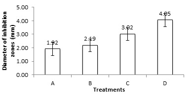
Based on the calculation of the diameter of the inhibition zones formed by the crude extract of mangrove snail meat against E. coli bacteria, with a positive control of amoxicillin at 150 ppm, the highest diameter observed was 14.42 mm, with a standard deviation of 0.51 mm. The negative control in this study used sterile distilled water, resulting in a diameter and standard deviation of 0 mm. Single-factor analysis (ANOVA) of the inhibition zone diameter formation revealed that the calculated F-value (119.14) was greater than the critical F-table value at a 1% significance level (3.70). Therefore, the hypothesis (H1) that the crude extract of mangrove snail meat (T. sulcata) at various concentrations effected the growth of E. coli was accepted. Since Fcalc > Ftable, it can be concluded that there is a significant difference in the concentration of T. sulcata crude extract affecting the growth of E. coli. Post hoc analysis using Tukey's Honestly Significant Difference (HSD) test indicated that concentration D had a significant difference, making it the most effective concentration in inhibiting the growth of E. coli with an average diameter of 4.05 mm.
The influence of concentration on the growth of E. coli was observed to be directly proportional, with an increase in extract concentration corresponding to an increase in the inhibition zone diameter (14). The formation of these inhibition zones is attributed to the presence of secondary metabolite compounds in the crude extract of T. sulcata (15). Phytochemical testing results showed that the crude extract of T. sulcata contained active compounds, including alkaloids, flavonoids, saponins, terpenoids, and phenols. These secondary metabolites exhibited antibacterial activity through various synergistic mechanisms, presumably by disrupting the structure and metabolic systems of the bacteria (16). The measurement of inhibition zone formation by T. sulcata crude extract, ranging from concentration A to D, against E. coli, with an average diameter ranging from 1.92 mm to 4.05 mm, falls into the category of weak antibacterial activity (17). Observations of inhibition zone formation at 2x24 h and 3x24 h showed a decrease compared to previous observations, indicating that all concentrations of T. sulcata crude extract have the potential as bacteriostatic antibiotics.
Conclusion
The composition of active compounds found in the crude extract of T. sulcata includes alkaloids (strong), flavonoids (weak), saponins (very strong), phenols (weak), and terpenoids (strong). The influence of concentration on the growth of E. coli is directly proportional, with increasing extract concentration corresponding to an increase in the diameter of the clear zones observed. The results of clear zone formation by T. sulcata meat extract, ranging from concentration A to D, against E. coli fall into the category of weak antibacterial activity. These findings indicate that the presence of strong saponins and terpenoids in the extract has a significant impact on inhibiting E. coli growth, albeit with a relatively weak antibacterial effect overall.
Declarations
Conflict of Interest
The author declares no conflicting interests.
Data Availability
The unpublished data is available upon request to the corresponding author.
Ethics Statement
Not applicable.
Funding Information
Not applicable.
References
- Barnes RSK. Interactions between benthic molluscs in a Sulawesi mangal, Indonesia: the cerithiid mud-creeper Cerithium coralium and potamidid mud-whelks, Terebralia spp. J Mar Biol Assoc United Kingdom. 2003 Jun 9;83(3):483–7.
- Sumarto Sumarto, Desmelati Desmelati, Dahlia Dahlia, Bustari Hasan MA. Penentuan senyawa bioaktif ekstrak daging siput bakau (Terebralia sulcata) dengan kromatografi lapis tipis (KLT). Berk Perikan Terubuk. 2011;39(2):85–96.
- Alimuddin Ali, Yusminah Hala and D. Penapisan dan karakterisasi parsial senyawa antimikroba dari siput bakau dan profil kromatografi lapis tipis fraksi aktif. Berk Penelit Hayati. 2006;12:63–8.
- Burrens NS, Clement JJ. Biomedical Potensial Marine Natural Product, Edited by Atawwa et al,(I), Phamaceutical and Bioactive Natural Product. New York and London: Plenum Press; 1993. 13–14 p.
- Abeysinghe P. Antibacterial activity of some medicinal mangroves against antibiotic resistant pathogenic bacteria. Indian J Pharm Sci. 2010;72(2):167.
- Van Giau V, An SSA, Hulme J. Recent advances in the treatment of pathogenic infections using antibiotics and nano-drug delivery vehicles. Drug Des Devel Ther. 2019 Jan;Volume 13:327–43.
- Kaper JB, Nataro JP, Harry L. T. Mobley. Pathogenic Escherichia coli. Nat Rev Microbiol. 2004;2:123–140.
- Köckerling E, Karrasch L, Schweitzer A, Razum O, Krause G. Public Health Research Resulting from One of the World’s Largest Outbreaks Caused by Entero-Hemorrhagic Escherichia coli in Germany 2011: A Review. Front Public Heal. 2017 Dec 11;5.
- Tribble DR. Antibiotic Therapy for Acute Watery Diarrhea and Dysentery. Mil Med. 2017 Sep;182(S2):17–25.
- Fair RJ, Tor Y. Antibiotics and Bacterial Resistance in the 21st Century. Perspect Medicin Chem. 2014 Jan 28;6:PMC.S14459.
- C Reygaert W. An overview of the antimicrobial resistance mechanisms of bacteria. AIMS Microbiol. 2018;4(3):482–501.
- AlSheikh HM Al, Sultan I, Kumar V, Rather IA, Al-Sheikh H, Tasleem Jan A, et al. Plant-Based Phytochemicals as Possible Alternative to Antibiotics in Combating Bacterial Drug Resistance. Antibiotics. 2020 Aug 4;9(8):480.
- Harborne JB. Metode Fitokia. Bandung: ITB; 1987.
- Gupta A, Jeyakumar E, Lawrence R. Strategic approach of multifaceted antibacterial mechanism of limonene traced in Escherichia coli. Sci Rep. 2021 Jul 5;11(1):13816.
- Setiaji J, Feliatra F, Teruna HY, Lukistyowati I, Suharman I, Muchlisin ZA, et al. Antibacterial activity in secondary metabolite extracts of heterotrophic bacteria against Vibrio alginolyticus, Aeromonas hydrophila, and Pseudomonas aeruginosa. F1000Research. 2020 Dec 21;9:1491.
- Allemailem K. Antimicrobial potential of naturally occurring bioactive secondary metabolites. J Pharm Bioallied Sci. 2021;13(2):155.
- Davis WW, Stout TR. Disc Plate Method of Microbiological Antibiotic Assay. Appl Microbiol. 1971 Oct;22(4):659–65.