Phytoconstituents and In Vitro Free Radical Scavenging Potential of n-Hexane and Aqueous Fractions of Cucurbita maxima and Leptadenia hastata
by Mubarak Muhammad Dahiru ★ , James Danga , Abdulhasib Oluwatobi Oni , Hesper Alex Zoaka, Rejoice Daniel Peter, Usanye Zira, Patience Christopher, Hauwa Yahaya Alkasim, Muhammad Zainab
Academic editor: Burak Kuzu
Sciences of Pharmacy 3(4): 193-202 (2024); https://doi.org/10.58920/sciphar0304265
This article is licensed under the Creative Commons Attribution (CC BY) 4.0 International License.
18 Jul 2024
25 Sep 2024
03 Nov 2024
11 Nov 2024
Abstract: The present study explored the phytoconstituents and radical scavenging activity of the respective n-hexane and aqueous fractions of Cucurbita maxima (CMHF and CMAF) and Leptadenia hastata (LHHF and LHAF) for potential application in oxidative stress-related ailments. The phytoconstituents were qualitatively determined and characterized using Fourier-transform Infrared (FTIR), while the antioxidant activity was determined in vitro. Alkaloids were present in only the aqueous fractions of C. maxima and L. hastata, while saponins, steroids, and flavonoids were detected in all the fractions. The FTIR revealed the presence of functional groups, including alcohols, sulfonates, alkenes, alkanes, amines, and aromatics in both plant fractions. The LHHF (35.53 ±2.11 ascorbic acid (AA) equivalent µg/mL) exhibited a significantly (p<0.05) higher total reducing power (TRP) than all the other fractions. The CMHF (69.11 ±2.56 AAE µg/mL) demonstrated a significantly (p<0.05) higher total antioxidant capacity (TAC) than all the other fractions. For the ferric thiocyanate (FTC) assay, the highest inhibition was exhibited by LHHF (79.78 ± 3.24%), significantly (p<0.05) higher than AA (26.46 ± 2.12%), CMHF (69.77 ± 3.16%), and CMAF (43.80 ± 2.12%). In the thiobarbituric acid assay, the lowest MDA concentration was exhibited by the CMHF (0.07 ±0.01 nmol/mL), significantly (p<0.05) lower than all the other fractions and ascorbic acid. Conclusively, the n-hexane fraction of both plants presents potential sources of novel antioxidant compounds with significant free radical scavenging and anti-lipid peroxidation activities, applicable in ailments linked to oxidative stress.
Keywords: AntioxidantsFunctional groupsLipid peroxidationMalonaldehyde concentration
Introduction
Oxidative stress (OS) defines a redox imbalance in a cell that is linked to several ailments, including cancer and diabetes. A state of redox imbalance is established in a cell when the number of free radicals released exceeds the capacity of the inherent antioxidant system of the body, notably in cancer and diabetes (1, 2). This is further exacerbated by the progression into an inflammatory statepromoted by the inflammatory cytokines (2, 3). Free radicals, including the reactive oxygen species (ROS) and reactive nitrogen species (RNS), are normal components of metabolic pathways and processes in a cell, such as phagocytosis, mitochondrial respiratory chain, and arachidonic acid metabolism (4). However, the antioxidant systems within the cell, such as glutathione peroxidase, catalase, and superoxide dismutase, neutralize and prevent their damaging effects on cellular components, including the DNA and membrane lipids (5). There are various free radicals, including peroxyl (ROO·), hydroxyl (·OH), alkoxy (-OR), nitric oxide (•NO), and superoxide (O₂•⁻). Additionally, hydrogen peroxide (H₂O₂), although not a radical, is often involved in oxidative processes (6). Once released without subsequent neutralization, these free radicals cause lipid peroxidation, protein modification, DNA fragmentation and damage, and cell death (1, 2). Thus, the need for free radical neutralization highlights the significant roles played by both endogenous and exogenous antioxidants in health and diseases.
Antioxidants, including the exogenous and their endogenous counterparts, are vital components of the cell, and their role is coupled to many pathways and cellular processes. Antioxidants act by donating or accepting electrons to free radicals, thereby neutralizing and preventing their associated effects (7). Exogenous sources of antioxidants entail compounds including phytochemicals with electron-donating capabilities, such as flavonoids, made up of the hydroxyl, carbonyl (oxo), and catechol groups within their structure. Moreover, the conjugated double bond within the flavonoid structure contributes to the antioxidant capabilities of these compounds (8, 9). Alkaloids, including allocryptopine, berberine, caffeine, and chelerythrine, exert their antioxidant properties by modulating pathways linked to OS (10). Allocryptopine modulates Akt/glycogen synthase kinase-3β (GSK-3β) signaling, significantly reducing OS-induced tau protein hyperphosphorylation. At the same time, berberine decreases endoplasmic reticulum (ER) stress and OS by suppressing ER PKR-like kinase (PERK)/eukaryotic translation initiation factor 2α (eIF2α) signalling-mediated beta-secretase 1 (BACE-1) translation (11, 12). Saponins, another class of phytochemicals, also exert their radical scavenging effects by modulating signaling cascade genes linked to OS (13). In a previous report, saponins were observed to upregulate antioxidant-related genes in human embryonic kidney 293 cells by activating nuclear factor erythroid 2-related factor 2 (13). Although these phytochemicals are attributed with antioxidant activities in cells, their radical scavenging potential presents a gap to explore in plant-based drugs for therapeutic application in OS-linked ailments.
Medicinal plants present a vast reserve of phytochemicals with different pharmacological activities, working individually or synergistically, including flavonoids, alkaloids, and saponins (14-18). In folkloric medicine, plant-based drugs prepared in different formulations are utilized against different ailments to achieve a desired therapeutic effect. L. hastata has been employed in traditional medicine to treat hypertension, erectile dysfunction, trypanosomiasis, diabetes mellitus, rhinitis (catarrh), skin diseases, and acute rhinopharyngitis (19, 20). C. maxima treats constipation, anemia, intestinal parasites, wounds, renal ailments, cataracts, and blood pressure (21, 22). The antioxidant activity of different plants, attributed to their phytoconstituents, was previously investigated via in vitro and in silico techniques (15, 16, 23-26). Some of these phytoconstituents were reported to be present in C. maxima and L. hastata, respectively. However, the extraction of these phytochemicals depends on the solvent employed during extraction. Moreover, the fractionation allows the partitioning of the phytochemicals based on solvent polarity. Previous studies focused on the in vitro antioxidant activities and characterization of the crude extracts of these plants via the gas-chromatography mass spectroscopy technique and 2,2-diphenyl-1-picrylhydrazyl assays, respectively (27, 28). However, in the present study, we characterized the phytochemicals and determined the antioxidant activity of the n-hexane and aqueous fractions of C. maxima and L. hastata seed and leaf, respectively, for potential application in oxidative stress-related ailments.
Methodology
Materials
C. maxima and L. hastata were collected from the bypass of Jimeta Area of Adamawa State, Nigeria. A Forest Technologist from the Forestry Technology Department of Adamawa State Polytechnic, Yola, identified the plant. A voucher specimen (No. ASP/FT/23/012) was deposited at the Department. The seed and leaf of C. maxima and L. hastata were air-dried and ground to powder using a blender to obtain the powdered sample.
Extraction
and Fractionation
Exactly 1 kg of the samples were separately macerated in 3 L of 75% (v/v) aqueous ethanol for 7 days in an air-tight container with daily agitation. This was followed by filtration using Whatman no. 1 filter paper to obtain the filtrate, which was concentrated to dryness with a rotary evaporator (Buchi Rotavapor R-200) under reduced temperature and pressure to obtain the crude extracts (72 and 165 g of C. maxima and L. hastata, respectively). Exactly 50 g of C. maxima crude extract was completely dissolved in 200 mL of distilled water and transferred to a 500 mL separating funnel. Then, an equal volume of n-hexane and vigorous shaking was added for complete partitioning and collection of the n-hexane layer. The aqueous layer was continuously washed with an equal volume of n-hexane until a clear n-hexane layer was obtained. The n-hexane and aqueous layers were dried via the same procedure for the crude extracts to obtain 19 and 29 g of the n-hexane (CMHF) and aqueous (CMAF) fractions of C. maxima, respectively. For the L. hastata, 100 g of the crude extract was subjected to the same treatment to obtain 44 and 62 g of the n-hexane (LHHF) and aqueous (LHAF) fractions, respectively.
Phytochemical
Detection
The method previously described by Evans (29) was used to detect the presence of alkaloids, saponins, steroids, glycosides, terpenoids, and flavonoids.
FTIR
Characterization
The functional group identification was carried out using Perkin-Elmer FTIR (series 100), according to the method described previously (30). The IR spectrum Table was used to identify the functional groups by interpreting and comparing the peaks.
Radical
Scavenging Potential
The radical scavenging potential of the extracts, including the total reducing power (TRP), total antioxidant capacity (TAC), ferric thiocyanate assay (FTC), and thiobarbituric acid assay (TBA) were determined as follows:
TRP
The TRP was determined based on the method described by Oyaizu (31). Briefly, 0.25 mL of 100 µg/mL of the sample was mixed with 0.625 mL of 0.2 M phosphate buffer (pH 6.6) and 0.625 mL of 1% (w/v) potassium ferrocyanide. The mixture was incubated for 50 min at 50°C, followed by adding 0.625 mL of 10% (w/v) trichloroacetic acid (TCA) and 10 min centrifugation at 3000 rpm. Precisely 1.8 mL of the upper layer was collected and mixed with an equal volume of distilled water. The absorbance was read at 700 nm against a blank (without sample) after adding 0.36 mL of 0.1% FeCl3. Moreover, varying concentrations (20-100 µg/mL) of ascorbic acid (AA) mixed with the reagents were used to obtain the AA calibration curve. The result was expressed as AA equivalent (AAE) in µg/mL of the mean of triplicate determinations.
TAC
The TAC was determined following the protocol Prieto et al. described (32). Briefly, 0.5 mL of 300 µg/mL of the sample was mixed with phosphomolybdate reagent. The mixture was capped and incubated for 10 min at 95°C. The absorbance of the sample was read at 695 nm after cooling against a blank (without the sample). Furthermore, varying concentrations (20-100 µg/mL) of AA mixed with the reagent were used to obtain the AA calibration curve. The result was expressed as AAE in µg/mL of the mean of triplicate determinations.
FTC
The FTC assay was used to determine the effect of the extract on the early stage of lipid peroxidation according to the protocol of Kikuzaki and Nakatani (33). Briefly, 4 mL of 1 mg/mL of the sample dissolved in absolute ethanol was mixed with 0.05 M pH 7 phosphate buffer and 3.9 mL distilled water, capped, and incubated in a dark oven at 40°C for 10 min. Exactly 0.1 mL of the solution was mixed with 0.1 mL of 30% (w/v) ammonium thiocyanate, 9.7 mL of 75% (v/v) ethanol, and 0.1 mL of 0.02 M ferrous chloride in 3.5% (v/v) HCl. A blank solution with only the reagents was used as a control, while AA was standard. The absorbance was read immediately and after every 24 h at 532 nm until the maximum absorbance of the control was observed. The percentage inhibition of lipid peroxidation was evaluated from Equation 1. The result was expressed as the mean of triplicate determination.
Equation 1
Where At = absorbance of sample, while Ac = absorbance of control.
TBA
The effect of the extracts on the late stage of lipid peroxidation (MDA formation) was determined as described by Kikuzaki and Nakatani (33). Briefly, 1 mL of the prepared sample in the FTC method was mixed with 2 mL each of 20% (w/v) TCA and 0.5% (w/v) TBA. The mixture was capped and incubated in a water bath for 20 min at 90°C, followed by centrifugation at 3000 rpm after cooling. The absorbance of the supernatant was read at 532 nm on the last of the FTC method. The MDA concentration was evaluated from Equation 2.
Equation 2
Where OD = absorbance of the sample, while EC = extinction coefficient.
Statistics
The results were evaluated using the Statistical Package for Social Sciences (SPSS) version 22 software and expressed as the mean of ± standard error of the mean of triplicate determinations (± SEM). The group difference was evaluated by One-way analysis of variance, followed by Tukey’s multiple comparison test.
Results
Phytochemical Detection
The phytoconstituents detected in the CMHF, LHHF, CMAF, and LHAF are shown in Table 1. Alkaloids were present in only the aqueous fractions of C. maxima and L. hastata, while saponins, steroids, and flavonoids were detected in all the fractions. Moreover, glycosides and terpenoids were absent in all the fractions.
Table 1. Phytochemicals detected in the n-hexane and aqueous fractions of C. maxima and L. hastata.
|
Phytochemicals |
CMHF |
LHHF |
CMAF |
LHAF |
|
Alkaloids |
- |
- |
+ |
+ |
|
Saponins |
+ |
+ |
+ |
+ |
|
Steroids |
+ |
+ |
+ |
+ |
|
Glycosides |
- |
- |
- |
- |
|
Terpenoids |
- |
- |
- |
- |
|
Flavonoids |
+ |
+ |
+ |
+ |
|
Note: (+) = present and (-) = absent |
||||

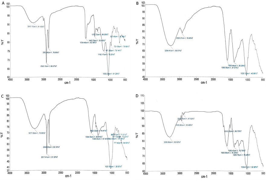
Figure 1. FTIR spectrum of n-hexane and aqueous fractions of C. maxima and L. hastata; (A) CMHF, (B) LHHF, (C) CMAF, and (D) LHAF.

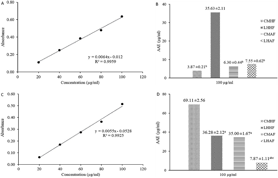
Figure 2. TRP and TAC of C. maxima and L. hastata; (A) AA TRP calibration curve, (B) TRP, (C) AA TAC calibration curve, and (D) TAC. Values with a, b, and c superscripts are significantly (p<0.05) lower than CMHF, LHHF, and CMAF, respectively.

Figure 3. Anti-lipid peroxidation potential of C. maxima and L. hastata; (A) FTC assay, (B) TBA assay. Values with a, b, c, d, and e superscripts are significantly (p<0.05) lower than CMHF, LHHF, CMAF, LHAF, and AA, respectively.
FTIR Characterization
The FTIR spectrum of the CMHF, LHHF, CMAH, and LHAF is presented in Figure 1, showing the absorption peaks. A total of 11 peaks were detected, including four in the group frequency region. For the CMHF, the first absorption peak was at 3317.17 cm-1, corresponding to the O-H stretching frequency of the alcohol compounds. The second (2923.10 cm-1) and third (2853.40 cm-1) peaks at the frequency regions correspond to the N-H stretching frequency of amine salt, while 1724.48 cm-1 corresponds to the C=O stretching of the α, β-unsaturated esters. In the fingerprint region, the peak at 1463.78 (C-H bending), 1377.72 (S=O stretching), 1192 (C-O stretching), 971.25 (C=H bend), 818.53 (C-H bending), and 721.05 cm-1 fingerprint regions corresponds to the alkane, sulfonates, ester, primary alcohol, alkene, disubstituted alkanes, aromatics, respectively. A total of 5 peaks were detected for the LHHF, including two in the group frequency region. The first absorption peak was at 3264.41 cm-1, corresponding to the O-H stretching frequency of the alcohol compounds. The second (2926.27 cm-1) peak at the frequency regions corresponds to the N-H stretching frequency of amine salt. In the fingerprint region, the peak at 1590.58 (N-H bending), 1398.44 (S=O stretching), and 1027.37 (C-N stretch) fingerprint regions correspond to sulfonates and amine groups, respectively.
A total of 11 peaks were detected for the CMAH, including three in the group frequency region. The first absorption peak was at 3277.22 cm-1, which corresponded to the O-H stretching frequency of the alcohol compounds. The second (2917.41 cm-1) and third (2849.25 cm-1) peaks at the frequency regions correspond to the N-H stretching frequency of amine salt. In the fingerprint region, 1594.59 (N-H bending), 1462.32 (C-H bending), 1398.00 (S=O stretching), 923.44 (C-H bend), 817.16 (C=C bending), and 777.40 cm-1 (C-H bend) fingerprint regions corresponds to the amine, alkane, sulfate, monosubstituted alkenes, trisubstituted alkenes, and ortho aromatics, respectively. A total of 8 peaks were detected for the LHAF, including four in the group frequency region. The first absorption peak was at 3288.06 cm-1, corresponding to the O-H stretching frequency of the alcohol compounds. The second (2935.82 cm-1) and third (2878.76 cm-1) peaks at the frequency regions correspond to the N-H stretching frequency of amine salt. In the fingerprint region, the peak at 1605.89 cm-1 corresponds to the C=C frequency of conjugated alkenes. The 1515.22 (N-O stretching), 1395.43 (S=O stretching), 1259.75 (C-N stretching), and 1027.37 (C-N stretch) fingerprint regions correspond to the nitrogen compound, sulfate, alkyl aryl ether, and amines, respectively.
Radical Scavenging Potential
TRP
The AA calibration curve and TRP of the CMHF, LHHF, CMAF, and LHAF are shown in Figure 2, depicting their AA equivalent total reducing power. The LHHF (35.53 ± 2.11 AAE µg/mL) exhibited a significantly (p<0.05) higher TRP than all the other fractions. Additionally, there was no significant (p>0.05) difference between the TRP of CMHF (3.87 ± 0.21 AAE µg/mL), CMAF (6.30 ± 0.44 AAE µg/mL), and LHAF (7.55 ± 0.62 AAE µg/mL) though the latter was slightly higher.
TAC
Figure 2 shows the AA calibration curve and TAC of the CMHF, LHHF, CMAF, and LHAF, revealing their AA equivalent total antioxidant capacity. The CMHF (69.11 ± 2.56 AAE µg/mL) demonstrated a significantly (p< 0.05) higher TAC than all the other fractions. Moreover, the LHAF (7.87 ± 1.11 AAE µg/mL) was significantly (p< 0.05) lower than the LHHF (36.28 ± 2.12 AAE µg/mL) and CMAF (35.00 ± 1.67 AAE µg/mL).
FTC
The anti-lipid peroxidation potential of CMHF, LHHF, CMAF, and LHAF is presented in Figure 3, showing the percentage inhibition. The highest inhibition was exhibited by the LHHF (79.78 ± 3.24%), significantly (p<0.05) higher than AA (26.46 ± 2.12%), CMHF (69.77 ± 3.16%), and CMAF (43.80 ± 2.12%). The CMHF (69.77 ± 3.16%) exhibited a significantly (p<0.05) higher inhibition than AA (26.46 ± 2.12%) and CMAF (43.80 ± 2.12%). Moreover, the LHAF (71.91 ± 2.66%) was significantly (p<0.05) higher than AA (26.46 ± 2.12%) and CMAF (43.80 ± 2.12%).
TBA
Figure 3 shows the anti-lipid peroxidation potential of CMHF, LHHF, CMAF, and LHAF, depicting the MDA concentration. The lowest MDA concentration was exhibited by the CMHF (0.07 ± 0.01 nmol/mL), significantly (p<0.05) lower than AA (0.37 ± 0.02 nmol/mL) and all the fractions. Moreover, the CMAH had a significantly (p<0.05) lower MDA concentration than AA (0.37 ± 0.02 nmol/mL), LHHF (0.36 ± 0.03 nmol/mL), and LHAF (0.42 ± 0.04 nmol/mL).
Discussion
Phytochemicals confer medicinal properties in plants and are implicated in the therapeutic effects of such plants. Different plants contain variable phytoconstituents, thus exhibiting different pharmacological effects. In our study, fractionation of the crude extract separated the phytochemicals based on polarity via the partitioning technique, which detected alkaloids in only the aqueous fractions. The polarity of solvents has been reported to influence the class of phytochemicals extracted, which also influences the pharmacological activities of the extracts, including their antioxidant potential (34). The phytochemicals detected in our study were previously implicated with antioxidant properties (10, 35, 36).
Moreover, the presence of these phytochemicals was previously reported (37). Mwangi et al. (37) reported similar results to the present study, detecting alkaloids, saponins, steroids, and flavonoids without terpenoids in C. maxima. In another study, Pal et al. (38) reported similar results to our study, detecting alkaloids and saponins in the aqueous seed extract of C. maxima. In their study, Omeh et al. (39) detected the presence of alkaloids, saponins, and flavonoids in L. hastata in addition to terpenoids and glycosides, slightly disagreeing with our report. However, the report of Maria and Olorukooba (40) on alkaloids, saponins, steroids, and flavonoids in L. hastata is concurrent with our result.
The radical scavenging capabilities of plant-based drugs are attributed to the presence of compounds and functional groups with electron-donating potential, neutralizing the free radicals generated from different pathways in cells. Additionally, these compounds act as ligands, modulating cell pathways and processes (11-13). Thus, detecting these functional groups in plants might offer antioxidant properties, including free radical scavenging effects. In our report, some of the functional groups detected include alkane, sulfonates, esters, alcohols, alkenes, disubstituted alkanes, aromatics, carboxylic acids, and amine salts which might be responsible for the radical scavenging of the plants. Chen et al. (41) reported the antioxidant activity of carboxylic acid and alcohol groups via a structure-activity relationship study, depicting enhanced antioxidant activity attributed to these groups. In another study, Fikriya and Cahyana (42) reported enhanced antioxidant activity of quinoline-4-carboxylic acid, synthesized through the Pfitzinger reaction via the reaction of a ketone with isatin. The result showed an increased antioxidant activity compared to the parent compound isatin, attributed to carboxylic acid and aromatic ring presence. Li et al. (43) reported a similar result with the presence of the carboxylic acid group in 1,8-Dihydroxyanthraquinone, leading to enhanced free radical scavenging compared to its analogs upon photoexcitation. The functional groups reported in our study were previously reported in other studies. Andronie et al. (44) reported the presence of alcohol, amine, alkane, and alkene groups in C. maxima seeds via the FTIR technique. Mwangi et al. (37) reported similar findings, identifying alcohol, amine, alkane, and alkene groups in C. maxima seeds. In another study, alcohol, amine, alkane, and alkene groups were detected in L. hastata leaf concurrently with our report (45).
The antioxidant activity is partly capable of scavenging free radicals by neutralizing and mitigating their effect on cellular components, including lipids and DNA (7, 46). This might serve as a first defensive strategy against free radicals in ailments associated with increased free radical generation, including diabetes, cancer, and neurodegenerative diseases. Thus, plant-based therapy to scavenge and neutralize the free radicals might serve beneficial effects. The ferric-reducing antioxidant capacity method is principled on reducing ferric to ferrous ions in the presence of the sample, yielding a directly proportional intense-blue color. Thus, the higher the absorbance, the higher the antioxidant capacity. A similar principle is followed for the total antioxidant capacity where Mo (VI) is reduced to Mo (V) by the antioxidant compounds present in the sample, which then forms a green phosphate/Mo(V) complex with maximum absorption at 695 nm. The principle behind this method relies on the ability of antioxidant molecules to donate electrons or hydrogen atoms to reactive species, thereby preventing oxidative damage. In our study, the superior total reducing power exhibited by the LHHF might be attributed to the presence of electron-donating compounds absent in the other fractions.
Interestingly, the CMHF showed superior total antioxidant capacity via the phosphomolybdate method. This variation might be attributed to the specificity of the antioxidant compounds present in the two fractions with preferences towards the ferric ions (Fe3+) or Mo (VI), demonstrating effectiveness to either assay (47). Further, AA exhibited superior TRP and TAC in our result, which correlates with the result reported by Kushawaha et al. (48) and Monica et al. (49). The TAC for the CMAF reported in our study was lower than the value reported by Kar et al., (28) and Tijjani et al., (50) for the crude aqueous extract of C. maxima which might be attributed to the absence of some compounds partitioned in the CMHF.
The FTC and TBA methods explore the early and end stages of lipid peroxidation, respectively, with the latter focused on inhibiting malonaldehyde (MDA) formation. MDA is often regarded as a biomarker of oxidative stress and the end product of lipid peroxidation, as increased lipid peroxidation leads to increased MDA formation. Although the LHHF showed the highest lipid peroxidation inhibition in our study, the CMHF exhibited the least MDA concentration, thus demonstrating superior activity against MDA formation. Kushawaha et al. (48) reported a comparable anti-lipid peroxidation activity of the aqueous extract of C. maxima seed with AA, which partially agrees with the result of the aqueous fraction in our study.
Conclusion
In the present study, the main objective was to explore the phytoconstituents and the free radical scavenging effect of n-hexane and aqueous fractions of C. maxima and L. hastata. The n-hexane fractions of both plants exhibited significant antioxidant activity, with L. hastata and C. maxima demonstrating superior total reducing power and total antioxidant capacity, respectively. Moreover, a similar observation was made for the anti-lipid peroxidation of the plants with superior activity exhibited by the n-hexane fractions of C. maxima and L. hastata via the FTC and TBA assays, respectively. These antioxidant activities might be attributed to the phytoconstituents with antioxidant functional groups identified in the FTIR analysis. Conclusively, both plants present a potential source of novel antioxidant compounds, with the n-hexane fractions demonstrating superior free radical scavenging and anti-lipid peroxidation activities. Thus, these plants can be applicable in the phytotherapy of ailments linked to oxidative stress, including diabetes, cancer, and aging-related diseases.
Abbreviations
AA = Ascorbic acid; CMAF = Cucurbita maxima Aqueous Fraction; CMHF = Cucurbita maxima n-hexane Fraction; FTC = Ferric Thiocyanate; FTIR = Fourier-transform Infrared; LHHF = Leptadenia hastata n-hexane Fraction; LHAF = Leptadenia hastata Aqueous Fraction; MDA = Malonaldehyde; OS = Oxidative Stress; ROS = Reactive Oxygen Species; RNS = Reactive Nitrogen Species; TAC = Total Antioxidant Capacity; TBA = Thiobarbituric Acid Assay; TRP = Total Reducing Power.
Declarations
Acknowledgment
The authors appreciate the contribution and institutional support of the Pharmaceutical Technology and Science Laboratory Technology Departments of Adamawa State Polytechnic, Yola.
Ethics Statement
Not applicable.
Data Availability
The unpublished data is available upon request to the corresponding author.
Funding Information
Not applicable.
Conflict of Interest
The authors declare no conflicting interest.
References
- Bhatti JS, Sehrawat A, Mishra J, Sidhu IS, Navik U, Khullar N, et al. Oxidative stress in the pathophysiology of type 2 diabetes and related complications: Current therapeutics strategies and future perspectives. Free Radical Biol Med. 2022;184:114-134.
- Hayes JD, Dinkova-Kostova AT, Tew KD. Oxidative Stress in Cancer. Cancer Cell. 2020;38(2):167-197.
- Yousef H, Khandoker AH, Feng SF, Helf C, Jelinek HF. Inflammation, oxidative stress and mitochondrial dysfunction in the progression of type II diabetes mellitus with coexisting hypertension. Front Endocrinol (Lausanne). 2023;14:1173402.
- Kıran TR, Otlu O, Karabulut AB. Oxidative stress and antioxidants in health and disease. Journal of Laboratory Medicine. 2023;47(1):1-11.
- Türkoğlu M, Pekmezci E, Sevinç H. Antioxidants and Antiaging. In: Alasalvar C, Shahidi F, Ho C-T, editors. Dietary Supplements with Antioxidant Activity: Understanding Mechanisms and Potential Health Benefits: The Royal Society of Chemistry; 2023. p. 0.
- Norma FS-S, Raúl S-C, Claudia V-C, Beatriz H-C. Antioxidant Compounds and Their Antioxidant Mechanism. In: Emad S, editor. Antioxidants. Rijeka: IntechOpen; 2019. p. Ch. 2.
- Sundaram Sanjay S, Shukla AK. Mechanism of antioxidant activity. Potential Therapeutic Applications of Nano-antioxidants: Springer; 2021. p. 83-99.
- Hassanpour SH, Doroudi A. Review of the antioxidant potential of flavonoids as a subgroup of polyphenols and partial substitute for synthetic antioxidants. Avicenna journal of phytomedicine. 2023;13(4):354.
- Speisky H, Shahidi F, Costa de Camargo A, Fuentes J. Revisiting the Oxidation of Flavonoids: Loss, Conservation or Enhancement of Their Antioxidant Properties. Antioxidants [Internet]. 2022; 11(1).
- Sirin S, Nigdelioglu Dolanbay S, Aslim B. Role of plant derived alkaloids as antioxidant agents for neurodegenerative diseases. Health Sciences Review. 2023;6:100071.
- Liang Y, Ye C, Chen Y, Chen Y, Diao S, Huang M. Berberine improves behavioral and cognitive deficits in a mouse model of alzheimer’s disease via regulation of β-amyloid production and endoplasmic reticulum stress. ACS Chem Neurosci. 2021;12(11):1894-1904.
- Xu Z, Adilijiang A, Wang W, You P, Lin D, Li X, et al. Arecoline attenuates memory impairment and demyelination in a cuprizone-induced mouse model of schizophrenia. Neuroreport. 2019;30(2):134-138.
- Khan MI, Karima G, Khan MZ, Shin JH, Kim JD. Correction: Khan et al. Therapeutic Effects of Saponins for the Prevention and Treatment of Cancer by Ameliorating Inflammation and Angiogenesis and Inducing Antioxidant and Apoptotic Effects in Human Cells (vol 23, pg, 10665, 2022). Int J Mol Sci. 2023;24(20: 15235).
- Dahiru MM. Recent advances in the therapeutic potential phytochemicals in managing diabetes. Journal of Clinical and Basic Research. 2023;7(1):13-20.
- Dahiru MM, Musa N. Phytochemical Profiling, Antioxidant, Antidiabetic, and ADMET Study of Diospyros mespiliformis Leaf, Hochst Ex A. Dc Ebenaceae. J Fac Pharm Ankara/Ankara Ecz Fak Derg. 2024;48(2):412-435.
- Dahiru MM, Alfa MB, Abubakar MA, Abdulllahi AP. Assessment of in silico antioxidant, anti-inflammatory, and antidiabetic activites of Ximenia americana L. Olacaceae. Advances in Medical, Pharmaceutical and Dental Research. 2024;4(1):1-13.
- Dahiru MM, Musa N, Abaka AM, Abubakar MA. Potential Antidiabetic Compounds from Anogeissus leiocarpus: Molecular Docking, Molecular Dynamic Simulation, and ADMET Studies. Borneo J Pharm. 2023;6(3):249-277.
- Dahıru MM, Musa N. GC-MS Analysis, Antioxidant, Antidiabetic Activity, and ADMET Study of Diospyros mespiliformis Hochst. Ex A. DC. Ebenaceae Stembark. Hacettepe University Journal of the Faculty of Pharmacy. 2024;44(3):198-219.
- Dambatta SH, Aliyu BS. A survey of major ethno medicinal plants of Kano north, Nigeria, their knowledge and uses by traditional healers. Bayero Journal of Pure and Applied Sciences. 2011;4(2):28-34.
- Yaro RS, Deeni YY, Abdullahi M, Bala I, Kawo AH. Medicinal Potential of Leptadenia hastata: A Review. Dutse Journal of Pure and Applied Sciences 2023;9(1b):15-21.
- Peter EL, Rumisha SF, Mashoto KO, Malebo HM. Ethno-medicinal knowledge and plants traditionally used to treat anemia in Tanzania: A cross sectional survey. J Ethnopharmacol. 2014;154(3):767-773.
- Salehi B, Capanoglu E, Adrar N, Catalkaya G, Shaheen S, Jaffer M, et al. Cucurbits Plants: A Key Emphasis to Its Pharmacological Potential. Molecules [Internet]. 2019; 24(10).
- Dahiru MM, Ahmadi H, Faruk MU, Aminu H, Hamman, Abreme GC. Phytochemical Analysis and Antioxidant Potential of Ethylacetate Extract of Tamarindus Indica (Tamarind) Leaves by Frap Assay. Journal of Fundamental and Applied Pharmaceutical Science. 2023;3(2):45-53.
- Dahiru MM, Nadro MS. Phytochemical Composition and Antioxidant Potential of Hyphaene thebaica Fruit. Borneo J Pharm. 2022;5(4):325-333.
- Dahiru MM, Umar AS, Muhammad M, Fari II, Musa ZY. Phytoconstituents, Fourier-Transform Infrared Characterization, and Antioxidant Potential of Ethyl Acetate Extract of Corchorus olitorius (Malvaceae). Sciences of Phytochemistry. 2024;3(1):1-10.
- Musa N, Dahiru MM, Badgal EB. Characterization, In Silico Antimalarial, Antiinflammatory, Antioxidant, and ADMET Assessment of Neonauclea excelsa Merr. Sciences of Pharmacy. 2024;3(2):92-107.
- Umaru IJ, Badruddin FA, Umaru H. Antioxidant Properties and Antibacterial Activities of Leptadenia Hastata Leaves Extracts on Staphylococcus Aureus. Drug Designing & Intellectual Properties International Journal. 2018.
- Kar S, Dutta S, Yasmin R. A comparative study on phytochemicals and antioxidant activity of different parts of pumpkin (Cucurbita maxima). Food Chemistry Advances. 2023;3:100505.
- Evans WC. Trease and Evans' pharmacognosy: Elsevier Health Sciences; 2009. 608 p.
- Dahiru MM, Umar AS, Muhammad M, Waziri AuA, Fari II, Musa ZY. Phytoconstituents, Fourier-Transform Infrared Characterization, and Antioxidant Potential of Ethyl Acetate Extract of Corchorus olitorius (Malvaceae). 2024;3(1):1-10.
- Oyaizu M. Studies on products of browning reaction antioxidative activities of products of browning reaction prepared from glucosamine. The Japanese journal of nutrition and dietetics. 1986;44(6):307-315.
- Prieto P, Pineda M, Aguilar M. Spectrophotometric quantitation of antioxidant capacity through the formation of a phosphomolybdenum complex: specific application to the determination of vitamin E. Anal Biochem. 1999;269(2):337-341.
- Kikuzaki H, Nakatani N. Antioxidant effects of some ginger constituents. J Food Sci. 1993;58(6):1407-1410.
- Wakeel A, Jan SA, Ullah I, Shinwari ZK, Xu M. Solvent polarity mediates phytochemical yield and antioxidant capacity of Isatis tinctoria. PeerJ. 2019;7:e7857.
- Cui Y, Liu B, Sun X, Li Z, Chen Y, Guo Z, et al. Protective effects of alfalfa saponins on oxidative stress-induced apoptotic cells. Food Funct. 2020;11(9):8133-8140.
- Messaadia L, Bekkar Y, Benamira M, Lahmar H. Predicting the antioxidant activity of some flavonoids of Arbutus plant: A theoretical approach. Chemical Physics Impact. 2020;1:100007.
- Mwangi JW, Kiragu D, Chaka B. Phytochemical screening, FTIR and GCMS analysis of Cucurbita pepo seeds cultivated in Kiambu county, Kenya. Heliyon. 2024;10(9):e30237.
- Pal P, Singh SB, Singh A. Determination of physicochemical properties, antioxidant constituents by high-performance thin-layer chromatography fingerprinting, and antioxidant activity of Cucurbita maxima seeds. Asian J Pharm Clin Res. 2018;11(3):280-283.
- Omeh I, Peter I, Maina V, Sandabe U, William A, Mshelia P, et al. Preliminary Study of the Contractile Effects of the Aqueous Extract of Leptadenia hastata Leaf (Pers Decne) on Rat Uterus. Journal of Veterinary Advances. 2016;6:1-15.
- Maria M, Olorukooba A. Phytochemical, Toxicological, Antinociceptive and Anti-inflammatory effect of Methanol Leaf Extract of Leptadeniahastata(PERS.) DECNE 1 2 3 4. Nigerian Journal of Pharmaceutical and Biomedical Research. 2018;3(2):170-175.
- Chen J, Yang J, Ma L, Li J, Shahzad N, Kim CK. Structure-antioxidant activity relationship of methoxy, phenolic hydroxyl, and carboxylic acid groups of phenolic acids. Sci Rep. 2020;10(1):2611.
- Fikriya SH, Cahyana AH. Study of Antioxidant Activity of the Derivatives of Quinoline-4-carboxylic Acids by the Modification of Isatin via Pfitzinger Reaction. Makara Journal of Science. 2023;27(2):160-164.
- Li M, Yang X, Zhao M, Shang C, Wang D, Li J, et al. Effects of carboxyl- and amino-groups on the antioxidant activity of hydroxyanthraquinones with ESIPT property: A theoretical study. J Mol Liq. 2023;376:121497.
- Andronie L, Pop I, Matei F, Coroian A, Rotaru A, Sobolu R, et al. Results obtained by investigating pumpkin (Cucurbita maxima L.) using FT-IR spectroscopy. 2022.
- Abdallah BM, Ali EM. Therapeutic Potential of Green Synthesized Gold Nanoparticles Using Extract of Leptadenia hastata against Invasive Pulmonary Aspergillosis. Journal of Fungi. 2022;8(5):442.
- Pisoschi AM, Pop A. The role of antioxidants in the chemistry of oxidative stress: A review. Eur J Med Chem. 2015;97:55-74.
- Christodoulou MC, Orellana Palacios JC, Hesami G, Jafarzadeh S, Lorenzo JM, Domínguez R, et al. Spectrophotometric methods for measurement of antioxidant activity in food and pharmaceuticals. Antioxidants. 2022;11(11):2213.
- Kushawaha D, Yadav M, Chatterji S, Maurya G, Rai A, Watal G. Free radical scavenging index of Cucurbita maxima seeds and their LIBS based antioxidant elemental profile. International Journal of Pharmacy and Pharmaceutical Sciences. 2016;8.
- Monica S, John S, Ramakrishnan M, Arumugam P. Evaluation of Antioxidant and Antidiabetic Potential of Crude Protein Extract of Pumpkin Seeds (Cucurbita maxima L.). Asian Journal of Biological and Life Sciences. 2020;9:42-47.
- Tijjani H, Matinja A, Yahya M, Aondofa E, Sani A. In vitro antioxidant and antidiarrheal activities of aqueous and n-hexane extracts of Cucurbita maxima seed in castor oil-induced diarrheal rats. Natural Resources for Human Health. 2022;2.

 ETFLIN
Notification


