
RESEARCH ARTICLE
Protective Effects of Java Plum (Syzygium cumini) Leaf Extract on Serum Biomarkers in Lead-Exposed Rats
Sciences of Pharmacy|Vol. 4, Issue 3, pp. 180-185 (2025)
CC BY 4.0-2025 Authors
Views
Downloads
Shares
Received
May 6, 2025Revised
Jun 10, 2025Accepted
Jul 15, 2025Published
Jul 25, 2025
Abstract
Lead (Pb), a heavy metal recognized as a hazardous environmental toxin, triggers oxidative stress by catalyzing oxidation reactions that produce free radicals, ultimately leading to cell death. Liver cell damage due to lead can decrease serum albumin levels, while kidney damage can increase serum uric acid levels. To counteract this oxidative stress, the intake of exogenous antioxidants is necessary. Java plum (Syzygium cumini) leaves are abundant in natural antioxidants, including flavonoids and phenolic compounds. The compounds possess antioxidant potential by scavenging free radicals. The purpose of this study was to determine the effects of an extract of S. cumini on serum albumin and uric acid in rats induced with lead acetate. All treatments lasted 30 days. Testing for serum albumin and uric acid was performed using the DiaSys method. One-way ANOVA followed by the Bonferroni post hoc test revealed significant differences (p < 0.05) in serum albumin levels among the groups: the normal group (4.13 g/dL), the positive control (2.86 g/dL), and the treatment group (3.93 g/dL). Similarly, significant differences were observed in serum uric acid levels: the normal group (1.47 mg/dL), the positive control (3.14 mg/dL), and the treatment group (1.64 mg/dL). These results indicate that S. cumini extract helps mitigate the adverse effects of lead exposure on serum albumin and uric acid levels in rats. A p-value of less than 0.05 indicates that the observed differences are statistically significant and unlikely due to chance.
Introduction
Reactive molecules comprise an atomic structure containing at least one unpaired electron. In high concentrations, these reactive and unstable molecules can either attack essential cellular components or generate even more reactive species that cause damage, such as to DNA or proteins. Over time, damage from reactive molecules has been linked to chronic degenerative diseases, such as diabetes mellitus and coronary heart disease. Reactive molecules can be produced endogenously or through various external sources. One significant exogenous source is the presence of heavy metals, including cadmium, chromium, copper, mercury, lead, and zinc, which contaminate the environment. Among these, lead ranks as one of the most toxic environmental pollutants (1).
However, research on the therapeutic use of Java plum (Syzygium cumini) extracts (SFE) as an antioxidant agent to mitigate lead-induced toxicity remains limited, especially in Indonesia, including regions such as West Sumatra. This research gap underscores the need for further studies examining the potential of Java plum extract to mitigate oxidative stress induced by lead exposure within local contexts.
Lead (Plumbum, Pb), a Group IVA heavy metal, is characterized by its bluish-white appearance, electrical conductivity, and resistance to acid and corrosion. It is widely used in various industries, including as an anti-knock agent in fuels, in battery production, as an anti-corrosive, in leaded glass, and as a battery cathode. The extensive use of lead in daily life increases the risk of human exposure (2). A study in a Chinese battery factory reported blood lead levels reaching 67.7 μg/dL, far exceeding the CDC’s acceptable limit of 10 μg/dL (3). Lead toxicity can affect multiple organs, such as the liver, kidneys, circulatory system, and heart, and promotes oxidative stress by increasing the production of reactive oxygen species (ROS) (4). These ROS can damage key biomolecules such as DNA, proteins, and enzymes, ultimately triggering cell death. When ROS levels exceed the body's antioxidant capacity, oxidative stress occurs.
Lead is primarily eliminated via the liver and kidneys, though only a small fraction is excreted in the urine. Consequently, it tends to accumulate in organs, where it may impair both structural and functional integrity (5). The liver and kidneys are of particular concern due to their roles in detoxification and excretion, respectively (6). Liver and kidney damage from lead exposure can be assessed using several biomarkers. Albumin is a key biochemical parameter that changes when the body is exposed to excessive lead levels. It is synthesized by hepatocytes in the liver and released into the bloodstream. In humans, serum albumin maintains plasma oncotic pressure and acts as a transport protein for various substances (7). A decrease in albumin levels may occur due to liver damage caused by lead, mainly through oxidative stress mechanisms (8). This aligns with findings from Offor et al. and Suradkar et al., who demonstrated that albumin levels are useful indicators of liver function (9, 10). Similarly, uric acid, which is primarily excreted by the kidneys (65–75%), can accumulate when renal function is impaired. Increased levels are associated with purine-rich diets, obesity, and lead-induced nephrotoxicity (11-13). Even modest increases in serum uric acid within the normal range have been linked to cardiovascular risk, metabolic syndrome, and kidney stones. Evidence shows that lead exposure can impair renal uric acid excretion, making serum uric acid a useful indicator of kidney function (14-17). Evidence shows that lead exposure can impair renal uric acid excretion, making serum uric acid a useful indicator of kidney function.
In the case of lead poisoning, the body’s endogenous antioxidant defenses are often insufficient. Therefore, exogenous antioxidants are needed to combat oxidative damage. One promising source is the Java plum (Syzygium cumini), known for its rich antioxidant profile and various therapeutic properties, including antibacterial, anti-inflammatory, gastroprotective, and antidiabetic activities (18, 19). Among all plant parts, leaves contain high concentrations of bioactive phytochemicals, including crategolic acid, betulinic acid, β-sitosterol, flavonoids (such as myricetin and quercetin), and polyphenols. These compounds are potent free radical scavengers and support endogenous antioxidants, such as vitamin C (20, 21). Research on the use of Syzygium cumini extract (SCE) to reduce oxidative stress induced by lead toxicity remains limited. Therefore, this study aims to investigate the effect of SCE administration on serum albumin and uric acid levels in rats exposed to lead acetate. The findings are expected to provide valuable insight into the antioxidant potential of SCE as an external supplement for mitigating the harmful effects of lead poisoning.
Experimental Section
Materials
The materials used in this study included 24 male white rats (Rattus norvegicus), aged 2–3 months and weighing 150–200 g, obtained from the Immunology Laboratory at the Faculty of Pharmacy, Universitas Andalas, Padang, Indonesia. The rats received standard feed and rice husk bedding from a certified local supplier. Lead acetate (Merck, Germany) was used to induce toxicity, and distilled water was produced in-house. Syzygium cumini leaves were collected from cultivated plants in Limau Manis, Pauh, Padang City, West Sumatra, with permission from the landowner and authenticated at the Herbarium ANDA, Universitas Andalas. The ethanol extract was prepared using 96% ethanol (Brataco, Indonesia). Other reagents included 0.9% saline (Otsuka, Indonesia), 70% alcohol (Brataco), and Betadine (Mundipharma, Indonesia), the bromocresol green (BCG) method with a commercial kit (Albumin FS, Version 21.04) from DiaSys Diagnostic Systems GmbH (Holzheim, Germany), and 2, 4, 6-Tribromo-3-hydroxybenzoic acid (TBHBA) (Sigma-Aldrich (Product No. T1642; ≥ 98% purity; Merck KGaA, Darmstadt, Germany). Biochemical analyses were performed using Diasys Albumin FS and Uric Acid FS kits (Diasys, Germany). Supporting materials included syringes (Terumo, Japan), microtubes (Axygen, USA), pipettes and tips (Eppendorf, Germany), and gauze (OneMed, Indonesia). The equipment used included a MicroLab 300 analyzer (Vital Scientific, Netherlands), a centrifuge (Hettich, Germany), and a vortex mixer (Thermo Fisher, USA).
Extract Preparation
The extraction of Syzygium cumini leaves took place at the Pharmacology Laboratory, Faculty of Pharmacy, Andalas University. Fresh Java plum leaves, weighing approximately 2.5 Kg (equivalent to approximately 1 kg dry weight), were thoroughly cleaned, cut into small pieces, and air-dried in the shade to prevent thermal degradation of the active compounds. Once dried, the leaves were ground into a coarse powder. Maceration was performed with 96% ethanol in a 1: 10 ratio for 3 days in a dark container at room temperature, followed by an additional 3 days of re-maceration to ensure complete extraction. The resulting liquid extract was then concentrated using a rotary evaporator at 40°C under vacuum, yielding about 48 grams of pure, viscous extract (22, 23)
In Vivo Test
Animal Preparation
This study obtained ethical approval with registration No.870/UN.16.2/KEP-FK/2022 from the Faculty of Medicine, Ministry of Education, Culture, Research, and Technology, Andalas University. A total of 24 healthy male rats (Rattus norvegicus) were randomly selected and allowed to acclimatize for one week under standard laboratory conditions, following the OECD Guideline (24). After acclimatization, rats (Rattus novergicus) that met the inclusion criteria (normal body weight 150-200 g, active physical condition, normal feeding and drinking behavior, and no visible signs of illness or injury) were randomly assigned to three groups (n = 8 per group) using a simple randomization method. The study included a normal group, a positive control group, and a treatment group. Throughout the experiment, rats were housed individually in cages maintained at room temperature (22–25°C), under a 12-hour light-dark cycle and shielded from direct sunlight. The cages were cleaned daily, and the animals had unrestricted access to food and water. Before the start of treatment, the body weight and physical condition (including activity, appetite, and drinking behavior) of each rat were closely monitored. Any sick or deceased rats during the acclimatization phase were replaced with new rats that met the inclusion criteria. The treatment was administered once daily for 28 consecutive days according to a predetermined schedule. Although the allocation of rats into groups was randomized, the outcome assessment was not blinded.
Sample Administration
The normal group was fed a standard diet without any additional treatments for 30 days. The negative control received an oral dose of lead acetate at 40 mg/kg body weight each morning for the duration of the study. Similarly, the treatment group was administered the same daily dose of lead acetate each morning, followed by SCE at 150 mg/kgBW in the afternoon, between 2:00 and 3:00 PM WIB (GMT+7), for 30 consecutive days (25, 26). Both the lead acetate solution and the SCE extract were administered via oral gavage. During the administration process, each rat was carefully restrained, and the gavage needle was gently inserted along the lateral edge of the palate to ensure the liquid passed into the esophagus.
Albumin Level Measurement
Serum albumin concentrations were determined using a photometric method provided by DiaSys Diagnostic Systems GmbH (Holzheim, Germany), employing bromocresol green (BCG) as the chromogenic agent. The analysis was conducted on a Micro Lab 300 spectrophotometer (Vital Scientific, Fieren, The Netherlands), with absorbance readings taken at a wavelength of 546 nm. The assay relies on the binding of BCG to serum albumin under mildly acidic conditions, producing a measurable color transition from yellow-green to blue-green. Before sample analysis, the Micro Lab 300 was subjected to routine daily maintenance to ensure optimal performance. Blood samples were collected by withdrawing 2 mL of venous blood from each rat, which was then transferred into centrifuge tubes and left undisturbed for approximately 5 min. The samples were centrifuged at 3000 rpm for 10 min to separate the serum, which was then aliquoted into appropriately labeled microtubes. For the assay, three types of reaction tubes were prepared: a blank (10 µL of distilled water), a standard (10 µL of calibration solution), and a sample (10 µL of serum). To each tube, 1000 µL of albumin reagent was added, followed by gentle mixing and incubation at room temperature (20–25 °C) for 10 min. Absorbance values were recorded at 546 nm to determine albumin concentrations.
Standard Curve Preparation
To generate a standard curve, a series of calibration standards with known albumin concentrations, prepared from the DiaSys-provided stock calibration solution, were measured using the same assay procedure. Each standard was mixed with 1000 µL of albumin reagent, incubated for 10 min, and the absorbance was recorded. The resulting absorbance values were plotted against their respective albumin concentrations to create a standard curve, which was used to interpolate the albumin concentrations in the test samples.
Uric Acid Level Measurement
Serum uric acid concentrations were assessed using an enzymatic photometric method from DiaSys Diagnostic Systems GmbH, which utilizes 2, 4, 6-tribromo-3-hydroxybenzoic acid (TBHBA) as a chromogen. The analysis was performed using a Micro Lab 300 spectrophotometer. In this method, uric acid is enzymatically oxidized to allantoin by uricase, producing hydrogen peroxide as a byproduct. The hydrogen peroxide then reacts with 4-aminoantipyrine and TBHBA to yield a red-colored quinoneimine compound, the intensity of which is proportional to the uric acid concentration and is measured at 546 nm.
Blood samples were obtained by collecting 2 mL of venous blood from each rat, followed by centrifugation to separate the serum. Two assay approaches were employed: the substrate start and sample start methods. For the substrate start method, 20 µL of serum was added to reaction tubes containing 1000 µL of Reagent 1. After thorough mixing, the tubes were incubated at room temperature (20–25 °C) for 5 min. Following this, 250 µL of Reagent 2 was added, and the tubes were mixed again before undergoing a second incubation, either for 30 min at room temperature or for 10 min at 37 °C. Absorbance was measured at 546 nm against a reagent blank within one minute of incubation completion.
In the sample start method, Reagents 1 and 2 were first combined in a 4:1 ratio to prepare the working reagent. Then, 1000 µL of this mixture was added to 20 µL of serum in labeled tubes. The mixture was incubated under the same conditions, and the absorbance was recorded at 546 nm against a blank within one minute of the final incubation.
Statistical Analysis
The data distribution was assessed for normality and homogeneity; normality assessment utilized the Shapiro-Wilk test. After confirming normality, a One-Way Analysis of Variance (ANOVA) was conducted to ascertain whether there were significant differences in serum albumin and uric acid levels across the three groups: normal group, positive control, and treatment. To explore pairwise differences, the Bonferroni method was applied for multiple comparisons for the derived post hoc test.
Results and Discussion
Figure 1A illustrates the mean serum albumin levels across the experimental groups. Rats in the positive control group, exposed to lead acetate only, exhibited the lowest average albumin level (2.86 g/dL), which was significantly lower than that of the normal group (4.13 g/dL) and the treatment group receiving 150 mg/kg BW of SCE (3.93 g/dL). This finding suggests that lead exposure leads to a significant decrease in albumin levels, indicating liver damage. In contrast, administration of SCE effectively prevented this decline, maintaining albumin levels close to those of the normal group.
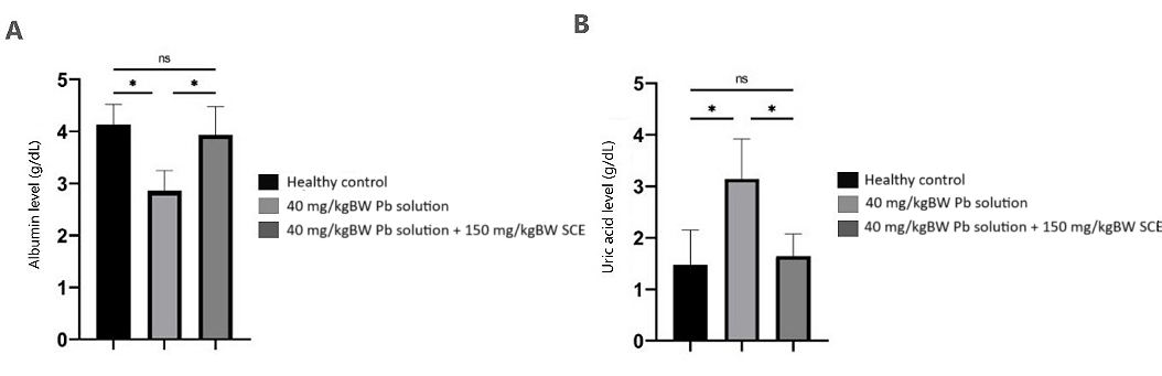
Statistical analysis confirmed significant differences in serum albumin and uric acid levels among the three groups (p < 0.05). The lower albumin levels in the lead-only group underscore the hepatotoxic effects of lead, consistent with previous studies that have linked lead exposure to impaired liver function and decreased albumin synthesis (9, 14, 7-29). The treatment group’s improved albumin levels suggest a hepatoprotective effect of SCE, likely due to its antioxidant properties, mitigating oxidative stress on hepatic cells.
Lead toxicity also severely impacts kidney function, which is reflected by changes in serum uric acid levels (see Figure 1B). The kidneys play a crucial role in filtering and excreting uric acid, so kidney impairment caused by lead results in elevated serum uric acid, a recognized biomarker for nephrotoxicity (30, 31). In this study, the positive control group exhibited significantly higher uric acid levels (3.14 mg/dL) compared to the normal group (1.47 mg/dL), indicating renal dysfunction associated with lead exposure.
The treatment group receiving SCE exhibited a notable reduction in uric acid levels (1.64 mg/dL) compared to the positive control, demonstrating the nephroprotective potential of SCE. This effect may be attributed to SCE’s ability to reduce oxidative stress in the kidneys by scavenging reactive oxygen species (ROS) and enhancing the activity of endogenous antioxidant enzymes, such as superoxide dismutase (SOD), catalase (CAT), and glutathione peroxidase (GPx) (32). Additionally, bioactive compounds in SCE, including flavonoids like quercetin, myricetin, and myricitrin, as well as phenolic acids such as ellagic and gallic acid, contribute to this protective effect by modulating inflammatory pathways (NF-κB and MAPK), inhibiting pro-inflammatory cytokines (TNF-α, IL-1β, IL-6), and preventing apoptosis in renal cells (23, 31-34).
The observed reduction in serum urea in the treatment group, as reported by Amriza et al. (2022), further supports the renal protective effects of SCE, which help restore kidney function impaired by lead toxicity (32). Lead accumulates in renal tubular epithelial cells, causing structural damage such as tubular atrophy and interstitial fibrosis, which progressively impair glomerular filtration and ultimately may lead to kidney failure (13, 28, 29, 33). The ability of SCE to counteract these pathological changes is likely linked to its multifaceted antioxidant and anti-inflammatory actions, which mitigate oxidative injury and maintain cellular homeostasis.
The administration of SCE in lead-exposed rats significantly improved both serum albumin and uric acid levels, indicating protection against lead-induced damage to the liver and kidneys. These findings suggest that SCE’s antioxidant and nephroprotective properties effectively restore normal physiological function, reducing oxidative stress and preventing tissue injury.
Conclusion
In conclusion, the study demonstrated that the average serum albumin and uric acid levels in the normal group of rats were 4.13 g/dL and 1.47 mg/dL, respectively. The positive control group exhibited mean serum albumin and uric acid levels of 2.86 g/dL and 3.14 mg/dL, respectively. Meanwhile, in the treatment group, the mean serum albumin and uric acid levels were 3.93 g/dL and 1.64 mg/dL, respectively. Significant differences were observed between the treatment and positive control groups, suggesting that the administration of SCE positively impacted serum albumin and uric acid levels. This positive effect is likely due to the antioxidant and protective properties of SCE, which help reduce oxidative stress and tissue damage caused by lead exposure. By protecting liver and kidney functions, SCE helps maintain albumin synthesis and promotes more efficient uric acid clearance from the bloodstream. Therefore, the administration of SCE helps restore normal serum albumin and uric acid levels in rats exposed to lead.
Abbreviations
SCE = Syzygium cumini Extract; ROS = Reactive Oxygen Species; MDA = Malondialdehyde; SGPT = Serum Glutamic Pyruvic Transaminase.
Declarations
Conflict of Interest
The authors declare no conflicting interest.
Data Availability
The unpublished data is available upon request to the corresponding author.
Ethics Statement
This study received ethical approval with number 870/UN.16.2/KEP-FK/2022 from the Faculty of Medicine, Ministry of Education, Culture, Research, and Technology, Andalas University
Funding Information
The author(s) declare that no financial support was received for the research, authorship, and/or publication of this article.
References
- Tumilaar SG, Hardianto A, Dohi H, Kurnia D. A Comprehensive Review of Free Radicals, Oxidative Stress, and Antioxidants: Overview, Clinical Applications, Global Perspectives, Future Directions, and Mechanisms of Antioxidant Activity of Flavonoid Compounds. Ahmed M, editor. J Chem [Internet]. 2024 May 31;2024:1–21.
- Nagaraju R, Kalahasthi R, Balachandar R, Bagepally BS. Association between lead exposure and DNA damage (genotoxicity): systematic review and meta-analysis. Arch Toxicol [Internet]. 2022 Nov 5;96(11):2899–2911.
- Collin MS, Venkatraman SK, Vijayakumar N, Kanimozhi V, Arbaaz SM, Stacey RGS, et al. Bioaccumulation of lead (Pb) and its effects on human: A review. Journal of Hazardous Materials Advances [Internet]. 2022 Aug;7:100094.
- Jomova K, Alomar SY, Nepovimova E, Kuca K, Valko M. Heavy metals: toxicity and human health effects. Arch Toxicol [Internet]. 2025 Jan 20;99(1):153–209.
- Jomova K, Raptova R, Alomar SY, Alwasel SH, Nepovimova E, Kuca K, et al. Reactive oxygen species, toxicity, oxidative stress, and antioxidants: chronic diseases and aging. Arch Toxicol [Internet]. 2023 Oct 19;97(10):2499–2574.
- Nakhaee S, Amirabadizadeh A, Brent J, Mehrpour O. Impact of chronic lead exposure on liver and kidney function and haematologic parameters. Basic Clin Pharmacol Toxicol [Internet]. 2019 May 18;124(5):621–628.
- Eruotor HO, Asiwe JN, Eruotor TM. Cymbopogon citratus protect against lead-induced suppression of haematological and tubuloglomerular functions as well as disruption of hepatocellular membranes in male Wistar rats. Journal of Trace Elements and Minerals [Internet]. 2023 Mar;3:100045.
- Wu N, Liu T, Tian M, Liu C, Ma S, Cao H, et al. Albumin, an interesting and functionally diverse protein, varies from ‘native’ to ‘effective’ (Review). Mol Med Rep [Internet]. 2023 Dec 13;29(2):24.
- Suradkar SG, Ghodasara DJ, Vihol P, Patel J, Jaiswal V, Prajapati KS. Haemato-biochemical alterations induced by lead acetate toxicity in Wistar rats. Vet World. 2009;2(11):429–431.
- Offor SJ, Mbagwu HOC, Orisakwe OE. Lead Induced Hepato-renal Damage in Male Albino Rats and Effects of Activated Charcoal. Front Pharmacol [Internet]. 2017 Mar 14;8.
- Fang X yu, Qi L wei, Chen H feng, Gao P, Zhang Q, Leng R xue, et al. The Interaction Between Dietary Fructose and Gut Microbiota in Hyperuricemia and Gout. Front Nutr [Internet]. 2022 Jun 22;9.
- Jiménez RT, Puig JG. Purine metabolism in the pathogenesis of hyperuricemia and inborn errors of purine metabolism associated with disease [Internet]. Gout and Other Crystal Arthropathies: Expert Consult: Online and Print. Elsevier; 2011. 36–50 p.
- Du L, Zong Y, Li H, Wang Q, Xie L, Yang B, et al. Hyperuricemia and its related diseases: mechanisms and advances in therapy. Signal Transduct Target Ther [Internet]. 2024 Aug 28;9(1):212.
- Maloberti A, Colombo V, Daus F, De Censi L, Abrignani MG, Temporelli PL, et al. Two still unanswered questions about uric acid and cardiovascular prevention: Is a specific uric acid cut-off needed? Is hypouricemic treatment able to reduce cardiovascular risk? Nutrition, Metabolism and Cardiovascular Diseases [Internet]. 2025 Mar;35(3):103792.
- Hussain M, Ghori MU, Aslam MN, Abbas S, Shafique M, Awan FR. Serum uric acid: an independent risk factor for cardiovascular disease in Pakistani Punjabi patients. BMC Cardiovasc Disord [Internet]. 2024 Oct 10;24(1):546.
- Crawley WT, Jungels CG, Stenmark KR, Fini MA. U-shaped association of uric acid to overall-cause mortality and its impact on clinical management of hyperuricemia. Redox Biol [Internet]. 2022 May;51:102271.
- Borghi C, Piani F. Uric Acid and Risk of Cardiovascular Disease: A Question of Start and Finish. Hypertension [Internet]. 2021 Nov;78(5):1219–1221.
- Rita RS, Sy E. Syzygium Cumini Leaves Extract from West Sumatra Indonesia Alleviate Oxidative Stress by Decreasing Malondialdehyde Level and Enhancing Catalase Activity in Rat Induced by Lead Acetate. Pharmacognosy Journal [Internet]. 2021 Nov 13;13(6):1408–1412.
- Anita Fajriyani, Anisah Fitriyani, Ridha Alisthipa Sephia, Hidayah H. The Potential of Jamblang Root or Java Plum (Syzygium cumini) in Medicinal Uses: A Systematic Review. Eureka Herba Indonesia [Internet]. 2023 Jun 6;4(2):216–220.
- Wongstitwilairoong N, Jermnark U, Paochoosak N, Limsivilai O, Chimnoi W, Yohannes Y, et al. Effect of Java plum (Syzygium cumini) leave extract and a silver nanoparticles synthesis on pathogens in skin diseases of dogs. Open Vet J [Internet]. 2024;14(10):2662.
- Rizvi MK, Rabail R, Munir S, Inam-Ur-Raheem M, Qayyum MMN, Kieliszek M, et al. Astounding Health Benefits of Jamun (Syzygium cumini) toward Metabolic Syndrome. Molecules [Internet]. 2022 Oct 24;27(21):7184.
- Das G, Nath R, Das Talukdar A, Ağagündüz D, Yilmaz B, Capasso R, et al. Major Bioactive Compounds from Java Plum Seeds: An Investigation of Its Extraction Procedures and Clinical Effects. Plants [Internet]. 2023 Mar 7;12(6):1214.
- Fatima CC, Agustini TW, Rianingsih L. The Effect of Java Plum Leaf Extract ( Syzygium Cumini ) on Vaname Shrimp Quality ( Litopenaeus Vannamei ) During Cold Storage. IOP Conf Ser Earth Environ Sci [Internet]. 2019 May 20;246:012018.
- OECD. OECD Guidelines for the Testing of Chemicals, Section 4: Health Effects – Test No. 407: Repeated Dose 28-day Oral Toxicity Study in Rodents [Internet]. Organisation for Economic Co-operation and Development.; 2008. (OECD Guidelines for the Testing of Chemicals, Section 4). Available from: https://www.oecd.org/en/publications/test-no-407-repeated-dose-28-day-oral-toxicity-study-in-rodents_9789264070684-en.html
- Ibrahim NM, Eweis EA, El-Beltagi HS, Abdel-Mobdy YE. The effect of lead acetate toxicity on experimental male albino rat. Biol Trace Elem Res. 2011;144(1–3):1120–1132.
- Rita RS, Sy E. Syzygium Cumini Leaves Extract from West Sumatra Indonesia Alleviate Oxidative Stress by Decreasing Malondialdehyde Level and Enhancing Catalase Activity in Rat Induced by Lead Acetate. Pharmacognosy Journal [Internet]. 2021 Nov 13;13(6):1408–1412.
- Faradita Diniyatuz Zahroh, Ramadhani Jaka Samudra, Rr Soenarnatalina Melaniani. Literature study: Comparative analysis of lead content in blood between spot workers, Caroseries industry workers, and metal smelting industry workers. World Journal of Advanced Research and Reviews. 2024;22(2):96–102.
- Rana MN, Tangpong J, Rahman MdM. Toxicodynamics of Lead, Cadmium, Mercury and Arsenic- induced kidney toxicity and treatment strategy: A mini review. Toxicol Rep [Internet]. 2018;5:704–713.
- Bruno CM, Pricoco G, Cantone D, Marino E, Bruno F. Tubular Handling of Uric Acid and Factors Influencing Its Renal Excretion: A Short Review. EMJ Nephrology. 2016;(July):92–7.
- Amin Rajizadeh M, Pourbabaki R. Oxidative Stress and Exposure to Metals. In 2024. Available from: https://www.intechopen.com/chapters/1190949
- Wang Y, Chen Y, Zhang X, Lu Y, Chen H. New insights in intestinal oxidative stress damage and the health intervention effects of nutrients: A review. J Funct Foods [Internet]. 2020 Dec;75:104248.
- Amriza MZ, Rita RS, Sy E. Java Plum (Syzygium cumini (L.) Skeels) Leaf Extract Lowers Serum Urea Levels in Lead-Acetate-Induced Rats. PHARMACY: Jurnal Farmasi Indonesia (Pharmaceutical Journal of Indonesia). 2022;19(2):274.
- Baki AE. The Effects of Lead Exposure on Serum Uric Acid and Hyperuricemia in Young Adult Workers: A Cross-sectional Controlled Study. Arch Rheumatol [Internet]. 2016 Mar 15;31(1):71–75.