Design and Screening of Tetracycline Antibiotics: An In Silico Approach
by Nahar Uddin Barbhuyan, Dubom Tayeng, Neelutpal Gogoi, Lima Patowary, Dipak Chetia, Malita Sarma Barthakur ★
Academic editor: James H. Zonthantluanga
Sciences of Phytochemistry 2(1): 7-13 (2023); https://doi.org/10.58920/sciphy02010008
This article is licensed under the Creative Commons Attribution (CC BY) 4.0 International License.
18 Nov 2022
08 Feb 2023
08 Feb 2023
11 Feb 2023
Abstract: A prominent class of broad-spectrum antibiotics known as tetracycline works by inhibiting the synthesis of proteins, which prevents the development of bacteria. Tetracycline resistance is typically attributed to one or more of the following causes: ribosomal binding site mutations, acquisition of mobile genetic elements carrying tetracycline-specific resistance genes, and/or chromosomal mutations that increase the expression of intrinsic resistance mechanisms. In this research, our objective is to virtually plan and conduct in-silico experiments to find tetracycline derivatives with inhibitory capability against tetracycline resistance protein. The tetracycline derivatives were screened using the Data Warrior, Discovery Studio, PyRx, and Swiss ADME web tools. Initially, 19 tetracycline derivatives were primarily screened for ADME and toxicity study followed by docking study. Among the tetracycline derivatives, C1, C11, C12, C14, C16, and C17 were found to be the potential drug-like molecules with binding energies of -8.9 kcal/mol, -8.4 kcal/mol, -8.5 kcal/mol, -7.7 kcal/mol, -7.7 kcal/mol, -8.6 kcal/mol respectively. In particular, C1 was predicted to have a better binding affinity towards the target protein than the others.
Keywords: In-silicoTetracyclineDrug resistanceTet R
Introduction
Tetracyclines are a significant group of broad-spectrum antibiotics that act by obstructing protein production to stop bacterial growth. Compounds of this broad family have bacteriostatic action and are used to treat a variety of bacterial infections, including Gram-positive and Gram-negative infections as well as diseases brought on by protozoan parasites and intracellular organisms (1). Tetracyclines have a hydronaphthacene nucleus and four linearly condensed benzene rings as their primary structural component.
Despite the notable success of penicillin, several industries, and academic institutions have been concerned with finding novel antibiotics synthesized by microorganisms by analyzing a large number of soil samples submitted from all over the world. Actinomycete bacteria were found to produce a yellow colony that had a remarkable inhibitory effect against a variety of pathogenic strains, including rickettsia and Gram-positive and Gram-negative bacteria. Aureomycin (syn. chlortetracycline) was the first tetracycline extracted from Streptomyces aureofaciens. With the isolation of Streptomyces rimosus by Pfizer, the antibiotics terramycin (syn. oxytetracycline) and aureomycin were obtained. Based on the chemical structure of Chlortetracycline by catalytic hydrogenation, tetracycline was discovered in 1953, with an improved pharmacokinetic profile. After discovering the first generation of tetracyclines (Chlortetracycline, oxytetracycline, and tetracycline), scientists have begun the development of newer tetracyclines with better pharmacokinetic properties, broader antimicrobial spectrum, and lower toxicity (2). The basic structure of tetracycline is given in Figure 1, substitution on the different positions can lead to different compounds. The classification of a different generations of tetracyclines has shown in Table 1.

Figure 1. General structure of tetracycline.
Table 1. Classification of the different generations of tetracyclines.
Generations | Obtaining method | Drugs |
First | Biosynthesis | Chlortetracycline, Oxytetracycline, Tetracycline, Demeclocycline |
Second | Semi-synthesis | Doxycycline, Minocycline, Lymecycline, Meclocycline, Methacycline, Rolitetracycline |
Third | Semi-synthesis Synthetic | Tigecycline, Omadacycline, Sarecycline Eravacycline |
For extended spectrum β-lactamase (ESBL)-producing Escherichia coli and Klebsiella species (spp.), the prevalence of tetracycline resistance was found to be 66.9% and 44.9%, respectively. For methicillin-resistant Staphylococcus aureus (MRSA) and Streptococcus pneumonia, the prevalence was 8.7% and 24.3%, respectively (3). Tetracycline resistance is typically attributed to one or more of the following factors: chromosomal mutations that increase the expression of intrinsic resistance mechanisms, the acquisition of mobile genetic elements carrying tetracycline-specific resistance genes, and/or ribosomal binding site mutations. Computational approaches in drug design, discovery, and development process gain very rapid exploration, implementation, and administration. Introducing a new drug into a market is a very complex, risky, and costly process in terms of money and manpower. For reducing time, cost, and risk-born factors, the computer aided drug design (CADD) method is widely used as a new drug design approach (4). There are mainly two types of approaches for drug design through CADDS: Structure-based drug design/direct approach and Ligand-based drug design/ indirect approach. Docking is a valuable approach for studying the many features connected to protein-ligand interactions. In the field of molecular modelling, it predicts the preferred orientation of one molecule to another when they are linked together to form a stable complex. In this study, different compounds have been designed based on the general structure of the tetracycline and performed basic in silico studies to find the best compound derivatives that can be analysed in resistant bacterial cells (5).
Materials and Methods
Retrieval of Target Protein
The target site is the regulator (Tet R) of a membrane-associated protein (Tet A) that exports the antibiotic out of the bacterial cell before it attaches to the ribosomes and inhibits polypeptide elongation. The X-Ray crystal structure of Tet R was downloaded from RCSB-PDB in PDB file format with a PDB ID of 2TCT. The crystal structure of the target protein is shown in Figure 2. The co-crystal inhibitor (CTC) was also identified and downloaded in SDF format.

Figure 2. Crystal structure of Tet repressor.
Preparation of Protein
The protein was prepared by the use of open-source BIOVIA Discovery Studio 2021 software (6). The water molecules and heteroatoms were also removed from the target protein. The active binding sites are defined with the ‘Define and edit binding site’ feature of the Discovery Studio software and the active site co-ordinates (x= 21.221424; y= 36.743182; z= 35.153182) were noted and saved for future use. Finally, polar hydrogen was added to the target protein and saved in PDB file format for future use.
Preparation of Compound Library
The structure of twenty compounds of tetracycline was prepared manually using Chem Draw Professional 16.0.0.82[68] software. The SMILES ID of these structures was also saved for future use. The compounds prepared with the software were saved in MDL SD File (*SDF) format.
Molecular Docking Simulation
Molecular docking simulation studies using Autodock Vina on PyRx 0.8 virtual screening platform were conducted to predict the binding affinity between the target proteins and the produced drugs (7). The prepared protein was inserted into the 3D scene in the software’s virtual platform and converted into PDBQT file format when processed into macromolecules (8). The downloaded proteins were reloaded. On enlarging Chain A of the corresponding protein, the amino acid sequence and the structure of the co-crystal ligand were made visible. To pinpoint precisely where the co-crystal inhibitors were located at the protein's binding site, the atoms of the co-crystal ligand were tagged. The pre-determined active binding site coordinates were utilized to modify the alignment of the 3D affinity grid box in the Vina search space of the PyRx 0.8 tool such that all of the amino acids are covered at the protein's active binding site. The default 3D affinity grid box size of 25 was maintained. And finally, MDSS was performed by the PyRx tool's predefined protocols (9).
Visualization and Analysis of Ligand Interactions
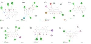
Using the Discovery Studio Visualizer Software, the 2D interaction of the compounds with the highest binding affinities to the protein was viewed. Additionally, the co-crystal-protein ligand interactions in two dimensions were seen. The compound was excluded from the investigation if it failed to create any regular hydrogen bonds with the active site residues.
ADMET Study
The ADME and toxicity prediction was done with Swiss ADME (10) and Data Warrior tools respectively. The SMILES ID of the compounds were entered in the required box and ADME and toxicity profiles (mutagenicity, carcinogenicity, reproductive toxicity, irritant) were generated.
Results
Features of the Target Protein and Tetracycline Derivatives
The RCSB-PDB website was used to get the crystal structure of protein chain A, shown in Figure 3. The protein is made up of Chain A, which is complexed with co-crystal inhibitor viz. CTC (Chlortetracycline). The 2D chemical structure of nineteen tetracycline derivatives is given in Figure 4.

Figure 3. Chain A of 2TCT (Tet Repressor) with co-crystal inhibitor.

Figure 4. 2D chemical structure of tetracycline derivatives.
Table 2. ADMET analysis of the compounds.
Compound | Molecular weight (g/mol) | H-bond acceptor | H-bond donor | iLogP | Lipinski violation | Toxicity | |||
Mutagenicity | Tumorigenicity | Reproductive | Irritant | ||||||
C1 | 463.41 | 11 | 6 | 1.62 | Yes | No | No | No | No |
C2 | 524.32 | 10 | 6 | 2.04 | No | No | No | No | No |
C3 | 470.43 | 11 | 6 | 0.68 | No | No | No | No | No |
C4 | 513.42 | 13 | 6 | 1.48 | No | No | No | No | No |
C5 | 487.46 | 11 | 6 | 1.20 | No | No | No | No | No |
C6 | 460.43 | 10 | 7 | 0.25 | No | No | No | No | No |
C7 | 474.46 | 10 | 7 | 1.37 | No | No | No | No | No |
C8 | 488.49 | 10 | 6 | 1.93 | No | No | No | No | No |
C9 | 461.42 | 11 | 7 | 1.59 | No | No | No | No | No |
C10 | 488.49 | 10 | 6 | 1.93 | No | No | No | No | No |
C11 | 459.45 | 10 | 6 | 2.03 | Yes | No | No | No | No |
C12 | 473.47 | 10 | 6 | 2.02 | Yes | No | No | No | No |
C13 | 459.45 | 10 | 6 | 2.03 | Yes | No | No | No | No |
C14 | 487.50 | 10 | 6 | 2.33 | Yes | No | No | No | No |
C15 | 501.53 | 10 | 6 | 1.42 | No | No | No | No | No |
C16 | 471.46 | 10 | 6 | 2.05 | Yes | No | No | No | No |
C17 | 469.44 | 10 | 6 | 1.85 | Yes | No | No | No | No |
C18 | 521.52 | 10 | 6 | 2.35 | No | No | No | No | No |
C19 | 469.44 | 10 | 6 | 1.85 | Yes | No | No | No | No |
ADMET Analysis
The ADME characteristics and toxicity of the compounds were established after the binding affinity of the tetracycline derivatives was established. Due to weak ADME qualities, substances that are active in in vitro environments may not be as active in vivo. Therefore, it is necessary to research a compound's bioavailability. The Lipinski Rule of Five, also referred to as Pfizer's Rule of Five or simply the Rule of Five is a general guideline used to assess a chemical compound's drug-likeness or to determine whether it possesses chemical and physical characteristics that would likely make it an orally active drug in humans.
Drugs are frequently taken off the market from clinical use due to toxicity. Many researchers rely on Data Warrior, a trustworthy program, to forecast the toxicity of substances. We conducted an in silico toxicity study, to ascertain the toxicity of the compounds. The ADME and toxicity study analysis is given in Table 2.
Molecular Docking Simulation
The molecular docking method enables us to define the behaviour of small molecules in the binding site of target proteins and to better understand basic biological processes by simulating the interaction between a small molecule and a protein at the atomic level. To evaluate the binding potential of ligands toward a protein, a docking study using the PyRx program can produce a binding affinity value (kcal/mol) for each ligand. Table 3 consists of a list of binding affinities of the compounds to the protein binding site.
Table 3. Binding affinities of the compounds with the protein binding site.
Sl. No. | Compounds | Binding affinities (-kcal/mol) |
1. | CTC (Co-crystal inhibitor) | 7.7 |
2. | C1 | 8.9 |
12. | C11 | 8.4 |
13. | C12 | 8.5 |
14. | C13 | 7.5 |
15. | C14 | 7.7 |
17. | C16 | 7.7 |
18. | C17 | 8.6 |
20. | C19 | 7.6 |
Visualization and Analysis of Ligand Interactions
The visualization of the 2D ligand interactions was performed using Discovery Studio Visualizer software, which provides detailed insights into the binding interactions between the ligands and the target protein. This software allows for a comprehensive analysis of molecular docking results, highlighting key interactions such as hydrogen bonds, hydrophobic interactions, and electrostatic forces.
The 2D ligand interaction images of the compounds with the TerA protein are presented in Figure 5, illustrating the specific binding sites and interaction patterns of each compound. Additionally, a summary of ligand interactions, detailing the specific amino acids involved in binding for each drug, is provided in Table 4. This data helps to better understand the molecular interactions that contribute to the binding affinity and potential effectiveness of these compounds as inhibitors or therapeutic agents.
Table 4. Summary of target-ligand interactions.
Compounds | Conventional Hydrogen bond | Other binding sites |
CTC (Co-crystal inhibitor) | HIS100 (2.70Å), HIS103 (2.15Å), HIS139 (2.23Å) | -- |
C1 | ASN82 (2.09Å), GLN116 (1.71Å), HIS100 (2.40Å), SER135 (1.78Å, 2.54Å) | THR103 (2.79Å), GLN109 (3.58Å) |
C11 | ASN82 (2.57Å), HIS100 (2.29Å), GLN116 (1.75Å), SER135 (1.73Å) | GLN109 (3.51Å) |
C12 | HIS64 (2.91Å), ASN82 (2.62Å, 2.72Å), HIS100 (2.92Å), THR103 (3.00Å), GLN116 (1.78Å, 2.49Å), SER135 (1.80Å) | GLN109 (3.65Å), HIS139 (4.86Å) |
C14 | ARG104 (4.12Å), HIS139 (2.03Å) | VAL113 (4.49Å), ILE134 (3.90Å) |
C16 | GLY102 (2.33Å), ARG104 (1.95Å), HIS139 (2.57Å) | GLY102 (3.44Å), VAL112 (4.27Å, 5.13Å), LEU117 (4.26Å), ILE134 (4.42Å) |
C17 | GLY102 (1.93Å), ARG104 (2.58Å), GLN116 (2.11Å), HIS139 (2.10Å) | PRO105 (5.43Å), VAL113 (4.27Å), LEU117 (4.27Å, 5.17Å), LEU131 (4.35Å), ILE134 (5.16Å) |
Discussions
A compound's ADMET characteristics relate to how it is absorbed, distributed, metabolized, excreted, and processed by and through the human body. To assess a drug's pharmacodynamic activity, it is crucial to consider ADMET, which makes up the pharmacokinetic profile of the drug molecule (11). The computational technique named molecular docking is used to find a ligand that will fit energetically and physically at the binding site of a protein. A stronger bond between a substance and a protein is indicated by a binding affinity value that is more negative (12). Low binding affinity values also suggest that protein-ligand binding requires less energy. The first pose is always regarded as the ideal pose since it has the highest binding affinity and the last pose has the lowest binding affinity to the target protein (13).
All the 19 derivatives were studied for ADMET analysis and found that only 8 compounds were shown to retain the Lipinski rule and no toxicity has been reported. Further, the screened 8 compounds were subjected to docking study and visualization of ligand interactions. In comparison to the binding affinity of the co-crystal inhibitor, 6 tetracycline derivatives have exhibited a greater affinity for chain A of the Tet R protein. These 6 variants include C1 with fluoro substituent at R4 position (-8.9 kcal/mol), C11 with methyl group at R4 (-8.4 kcal/mol), C12 with ethyl group at R4 (-8.5 kcal/mol), C14 with isopropyl substituent at R4 (-7.7 kcal/mol), C16 with ethenyl substituent at R4 (-7.7 kcal/mol), C17 with ethynyl substituent at R4 (-8.6 kcal/mol). These substances are further studied for the visualization of ligand interactions. It can be seen from the ligand interactions in figure 4 and table 5 that all 6 compounds have formed traditional H-bonds with various amino acids in the active site of the Tet R protein. Among these 6 tetracycline derivatives, C1 is found to have a better binding affinity towards the target protein.
Researchers now apply to trim methods that differ from the traditional approaches used to examine synthetic medicines. In silico techniques like molecular docking and molecular dynamics simulations have been employed more and more in drug discovery research to locate promising synthetic compounds for the treatment of various ailments. But certain medications have poor oral bioavailability. To address the issues with drug bioavailability, many researchers have adopted novel drug delivery strategies. Scientific developments have led to the use of novel techniques in drug discovery programs by pharmaceutical researchers (14).
Conclusions
The results of the current in silico analysis indicate that six compounds can inhibit the Tet R protein of the tetracycline resistance element. Compared to the co-crystal inhibitor, they have shown a greater binding affinity for the Tet R's active binding site. Among the six compounds, Compound 1 (Fluorine at 7th position) has shown a better binding affinity towards the target protein. Additionally, these molecules have a favorable ADME profile and no toxicity. To effectively evaluate the compound's inhibitory potential against Tet R protein of the tetracycline resistance element, however, additional molecular dynamic simulation study and (in vitro/in vivo) research is required as the current work is limited to only molecular docking and ADMET in silico model.
Declarations
Acknowledgment
Dubom Tayeng gratefully acknowledges the University Grant Commission and the Ministry of Tribal Affairs, Government of India for providing a fellowship (Award No.: 202021-NFST-ARU-02761) to support his Ph.D. research work.
Ethics Statement
Not applicable.
Data Availability
The unpublished data is available upon request to the corresponding author.
Funding Information
Not applicable.
Conflict of Interest
The authors declare no conflicting interest.

 ETFLIN
Notification


