An In Vitro Assessment of the Antioxidant Activity of Detarium microcarpum Guill. & Perr. Fabaceae
by Mubarak Muhammad Dahiru ★ , Abdulhasib Oluwatobi Oni, James Danga , Aisha Alfa Alhaji, Faith Jonah, Alkasim Yahaya Hauwa , Zainab Muhammad
Academic editor: Samir Chtita
Sciences of Phytochemistry 3(2): 114-122 (2024); https://doi.org/10.58920/sciphy0302267
This article is licensed under the Creative Commons Attribution (CC BY) 4.0 International License.
24 Jul 2024
20 Oct 2024
18 Nov 2024
27 Nov 2024
Abstract: Medicinal plants are regarded as important sources of exogenous antioxidants due to their phytoconstituents’ free radical scavenging potential. The present study explores the phytoconstituents and antioxidant activity of n-hexane (HFDM) and chloroform (CFDM) fractions of Detarium microcarpum for potential use in the phytotherapy of oxidative stress-linked ailments. The phytoconstituents were qualitatively determined, while the antioxidant activity was determined by in vitro assays. Alkaloids, saponins, steroids, and flavonoids were detected in both fractions, while glycosides and terpenoids were absent. The HFDM (55.40 ± 2.66 AAE µg/mL) showed a significantly higher total antioxidant capacity than the CFDM (34.84 ± 1.22 AAE µg/mL, p<0.05) at the tested concentration (100 µg/mL) while the CFDM (57.84 ± 2.16 AAE µg/mL) exhibited a significantly higher ferric reducing antioxidant power than the HFDM (46.11 ± 1.91 AAE µg/mL, p<0.05) at the tested concentration (100 µg/mL). In the ferric thiocyanate assay, there was no significant (p>0.05) difference between the HFDM (65.59 ± 2.32%) and CFDM (77.42 ± 2.95%). However, both fractions exhibited significantly higher percentage inhibition than ascorbic acid (9.68 ± 5.41%, p<0.05). Moreover, the HFDM (0.21 ± 0.01 nmol/mL) exhibited a significantly lower MDA concentration than the CFDM (0.66 ± 0.06 nmol/mL) and AA (0.46 ± 0.02 nmol/mL). Additionally, ascorbic acid (0.46 ± 0.02 nmol/mL) showed a significantly lower MDA concentration than CFDM (0.66 ± 0.06 nmol/mL). The n-hexane and chloroform fractions of the plants showed promising antioxidant potential, which might be attributed to the identified phytochemicals that have potential applications in the phytotherapy of oxidative stress-linked diseases.
Keywords: Free radicalsLipid peroxidationOxidative stressPhytochemical compositionPhytotherapy
Introduction
Antioxidants are regarded as vital cellular components, widely recognized for their key role in maintaining redox balance and mitigating the damaging effects of free radicals, including reactive oxygen species (ROS) and reactive nitrogen species (RNS), generated during normal cellular processes (1-6). The cell is inhabited by its inherent antioxidant enzymes, including glutathione, superoxide dismutase, and catalase, in addition to the exogenous antioxidant compounds such as ascorbic acid and tocopherol (3-5, 7). Antioxidants usually neutralize free radicals generated during normal cellular processes by donating electrons in some instances or by modulating pathways linked to the cellular processes (1, 3, 4, 8). However, in ailments associated with increased free radical generation, including cancer and diabetes, a redox imbalance is established, leading to oxidative stress and depletion of the antioxidants (6, 9-12). Oxidative stress is a major component of many ailments, subsequently leading to inflammatory responses and promoting tissue damage, which might lead to fatal consequences (13-16). In diabetes, persistent hyperglycemia promotes oxidative stress via mitochondrial dysfunction, endoplasmic reticulum stress, chronic low-grade inflammation, and lipotoxicity, further exacerbating the condition and leading to microvascular and macrovascular complications (13, 17-19). A similar event occurs in cancer, including mitochondrial dysfunction and endoplasmic reticulum stress. However, there is an increased activity of ROS-generating enzymes, including NADPH oxidase, cyclooxygenase (Cox), and xanthine oxidase in the cancer cells in addition to the Warburg effect, characterized by increased glucose metabolism even in the presence of oxygen (14, 16). Antioxidants play a vital role in health and diseases; however, their depletion in disease state might require augmentations from endogenous sources such as medicinal plants to mitigate oxidative stress.
Medicinal plants present a reservoir of plant-based drugs and natural products with varying pharmacological activities (20-28). Moreover, their abundance, affordability, and accessibility might provide an alternative to managing different ailments, including oxidative stress. In normal cellular conditions, ROS are generated as part of normal metabolic activities within the cell and subsequently neutralized to mitigate their damaging effects within the cell (29). However, the loss of cellular homeostasis due to the overproduction of ROS leads to harmful impacts on macromolecules, manifesting in ailments such as cancer and diabetes. Furthermore, this imbalance, coupled with the depletion of the inherent antioxidant system, further exacerbates these conditions, thus requiring an exogenous supply of antioxidants. In some reports, in vitro techniques were employed to determine the free radical scavenging activities of the plants, while other reports express the antioxidant effects in silico. In the in vitro techniques, including the ferric-reducing antioxidant assay and phosphomolybdate assay, different plant extracts and fractions are used in the presence of ferric and molybdate ions to explore the radical scavenging potential of the plants (24, 30-34). The in silico technique determines the modulative effects of compounds identified in the plants on the different proteins and enzymes associated with generating free radicals (28, 32, 33, 35). The therapeutic effects of medicinal plants are attributed to their phytochemical components, which they produce for plant growth and defence against predators and pathogens (21, 22, 26, 36-38). Phytochemicals, including saponins and flavonoids, are linked to different antioxidant activities via different mechanisms of action, such as direct free radical neutralization by donating electrons and modulating the activities of different cellular processes linked to free radical generation (39, 40).
D. microcarpum is a tree native to drier parts of Africa, including Nigeria, Sudan, and Senegal, growing up to 36 m with a thick and big crown, depending on the amount of rainfall in the area (41, 42). In folkloric medicine, different parts of the plant are prepared in different formulations, including decoction, infusion, and maceration, and they are employed in treating infections, fever, menstrual pains, and heart diseases (43). Different parts of the plant are documented to contain different phytochemicals with pharmacological activities (44, 45), thus finding their way into different forms of phytotherapy. Additionally, some fatty acids, including linoleic, oleic, and palmitic acids, were previously identified in the plant and linked to potential antimicrobial activities of the plant (46, 47). The plant was previously linked to pharmacological activities, including antimicrobial, antiproliferative, antiplasmodial, analgesic, and anti-inflammatory (48-50, 45). Although the antioxidant activities of different parts of the plants were previously reported and attributed to their phytochemical components (51-53), there are still unexplored gaps in the quest to harness the full antioxidant potential of the plant. In the present study, the n-hexane and chloroform stembark fractions of D. microcarpum were assessed for their phytochemical composition and free radical scavenging activity for potential application as antioxidants in oxidative stress-linked ailments.
Methodology
Materials
A sample of D. microcarpum was obtained from the Girei Local Government Area of Adamawa State, which was identified and authenticated by a Forest Technologist in the Forestry Technology Department of Adamawa State Polytechnic Yola. The sample was dried under shade, ground to powder, and stored till further use.
Extraction
and Fractionation
The crude D. microcarpum extract was obtained by macerating the plant powder (one kilogram) in 75% (v/v) ethanol for 7 days with frequent agitation, followed by filtration with a Whatman No.1 filter paper to obtain the filtrate. The filtrate was dried over a rotary evaporator (Buchi Rotavapor R-200) under reduced pressure at 40°C to obtain 248 g of the crude extract. The crude extract was further subjected to fraction via solvent partitioning technique to get the n-hexane and chloroform fractions. Exactly 50 g of the crude extract was dissolved in 100 mL of distilled water and transferred to a separating funnel. An equal volume of n-hexane was added, shaken vigorously, and allowed to stand for separation. The n-hexane layer was collected, and the process was repeated until a clear n-hexane layer was observed. Furthermore, an equal volume of chloroform was added to the aqueous fraction and subjected to the same treatment as the n-hexane fraction. The n-hexane and chloroform layers were collected separately and dried via the same procedure for the crude extract to obtain the n-hexane (HFDM) and chloroform (CFDM) fractions, respectively.
Phytochemical
Identification
The methods previously described by Evans (54) were adopted to detect the presence of alkaloids, saponins, steroids, glycosides, terpenoids, and flavonoids.
Alkaloids
Exactly 2 mL of 10% HCl was added to 2 mL of the extract, followed by the addition of 2 mL of Meyer’s reagent. The formation of an orange precipitate indicated a positive result.
Saponins
Exactly 2 mL of distilled water was added to 2 mL of the extract, followed by 5 minutes of agitation. The appearance of a foam layer indicated a positive result.
Steroids
Exactly 10 mL of chloroform was added to 2 mL of the extract, followed by the addition of 10 mL of concentrated sulphuric acid by the side of the test tube. The formation of a reddish upper layer and yellow sulphuric acid layer with green fluorescence indicated a positive result.
Glycosides
Exactly 2 mL of acetic acid was added to 2 mL of the extract. The mixture was cooled in a cold water bath, and then 2 mL of concentrated H2SO4 was added. Color development from blue to bluish-green indicated the presence of glycosides.
Terpenoids
Exactly 2 mL of chloroform and 1 mL of concentrated sulphuric acid were carefully added to 2 mL of the extract to form a layer. A clear upper and lower layer with a reddish-brown interphase indicated a positive result.
Flavonoids
Exactly 2 mL of 10% sodium hydroxide was added to 2 mL of the extract. The formation of a yellow color, which turned colorless upon the addition of 2 mL of dilute hydrochloric acid, indicated a positive result.
Antioxidant
Activity
Different methods were adopted to determine the in vitro free radical scavenging activity and anti-lipid peroxidation potential of the HFDM and CFDM.
Total
Antioxidant Capacity
The TAC was determined by adopting the method previously described by Prieto et al. (55). Exactly 0.5 mL of distilled-water dissolved sample (300 µg/mL) was mixed with 2 mL of phosphomolybdate reagent and capped, followed by 10 minutes of incubation at 95°C. The sample absorbance was read using a UV-Viz spectrophotometer (Model V1000) at 695 nm against a blank solution of phosphomolybdate reagent and distilled water subjected to the same treatment as the sample. Moreover, different concentrations (20 – 100 µg/mL) of ascorbic acid (AA) were used to determine the AA calibration curve. The TAC was expressed as AA equivalent (AAE) in µg/mL of triplicate determinations.
Ferric
Reducing Antioxidant Power
The FRAP was determined as described previously by Oyaizu (56). Exactly 0.25 mL distilled-water dissolved sample (100 µg/mL) was mixed with 0.625 mL of 1% potassium ferrocyanide and 0.625 mL of 0.2 M pH 6.6 phosphate buffer, followed by 20 minutes incubation at 95°C. Furthermore, 0.625 mL of 10% trichloro acetic acid (TCA) was added, followed by 20 minutes of centrifugation at 3000 rpm and collection of 1.8 mL of the upper layer, which was mixed with an equal volume of distilled water and 0.36 mL of 0.1% FeCl3. The absorbance was read using a UV-Viz spectrophotometer (Model V1000) at 700 nm against a blank solution of reagents without the sample. The AA calibration curve was derived from different concentrations (20-100 µg/mL) of AA, and the result was expressed as AAE µg/mL of triplicate determinations.
Ferric
Thiocyanate Assay
The anti-lipid peroxidation potential of the sample was determined by adopting the method of Kikuzaki et al. (57) via the FTC assay. Precisely 4 mL (1 mg/mL) and 4.1 mL of the sample and 2.25% (v/v) linoleic acid prepared in absolute ethanol were combined with 3.9 mL pH 7 phosphate buffer and 3.9 mL distilled water. This was followed by capping and 10 minutes of dark oven incubation at 40°C. Furthermore, 0.1 mL of the mixture was mixed with 9.7 mL of 75% ethanol and 0.1 mL of 30% ammonium thiocyanate, followed by the addition of 0.1 mL of 0.02 M ferrous chloride prepared in 3.5% HCl. The initial absorbance of the mixture was read using a UV-Viz spectrophotometer (Model V1000) at 532 nm three minutes after the addition of ferrous chloride. Moreover, a solution of the reagents without the sample was used as a control, while AA was standard. The percentage inhibition was determined according to Equation 1 and presented as a mean of triplicate determinations.
Equation 1
Where At = absorbance of sample and while Ac = Absorbance of control.
Thiobarbituric
Acid Assay
The procedure previously described by Kikuzaki et al. (57) was adopted to further determine the anti-lipid peroxidation of the sample. One milliliter of the sample prepared in the FTC method was mixed with two milliliters of each of 20% TCA and 0.67% TBA, followed by capping and 10 minutes of incubation at 90 °C. The mixture was centrifuged at 3000 rpm for 20 minutes after cooling. The absorbance was read using a UV-Viz spectrophotometer (Model V1000) at 532 nm. Furthermore, a solution of the reagents without the sample is used as blank, while AA is standard. The malonaldehyde (MDA) concentration was determined from the extinction coefficient 156 Mm-1 cm-1 using Equation 2.
Equation 2
Where At = absorbance of the sample, EC = extinction coefficient, and Vs = sample volume.
Statistical Analysis
The results obtained in the present study were expressed as the mean of ± standard error of triplicate determinations (±SEM). Moreover, the difference among the groups was analyzed with Statistical Package for the Social Sciences (SPSS) version 22 Software using an independent-sample T-test at p<0.05 significance level.
Result
and Discussion
Phytochemical Identification
The phytochemical components detected in D. microcarpum are listed in Table 1. Both the HFDM and CFDM showed the presence of alkaloids, saponins, steroids, and flavonoids with the absence of glycosides and terpenoids. In our study, four classes of phytochemicals were identified in both fractions of D. microcarpum, including alkaloids, saponins, steroids, and flavonoids. The detection of similar phytochemicals in the HFDM and CFDM might be attributed to the polarity of the fractionating solvents, which isn’t surprising, considering the polarity range of the solvents (58-60). Furthermore, the identified phytochemicals were previously detected. Dahiru et al. (45) reported similar results, detecting the presence of alkaloids, saponins, and flavonoids in D. microcarpum with the absence of steroids. Sanusi et al. (50) reported the presence of alkaloids, saponins, steroids, glycosides, and flavonoids, partially agreeing with our results. In another study, alkaloids, flavonoids, steroids, and saponins were reported, which agrees with our report (61). Additionally, the phytochemicals reported in our study were previously attributed to different pharmacological activities, including antioxidant activities.
Table 1. Phytochemical Components of D. microcarpum.
|
No. |
Phytochemical |
HFDM |
CFDM |
|
1 |
Alkaloids |
+ |
+ |
|
2 |
Saponins |
+ |
+ |
|
3 |
Steroids |
+ |
+ |
|
4 |
Glycosides |
- |
- |
|
5 |
Terpenoids |
- |
- |
|
6 |
Flavonoids |
+ |
+ |
|
Note: (+) = present, (-) = absent |
|||
Alkaloids exert antioxidant activity against oxidative stress via modulation of pathways and enzymes linked to oxidative stress (62). Uthman et al. (63) reported concentration-dependent in vitro antioxidant activities of alkaloids and flavonoids from Kalanchoe pinnata, attributed to the identified compounds in the plant. In another report, alkaloid compounds isolated from Litsea cubeba demonstrated strong antioxidant activity in vitro which was linked to the functional groups present in the compounds (64). Lokesh et al. (65) reported the antioxidant activity of crude saponins from Abutilon indicum leaves with up to 96.16% free radical scavenging activity at 2.5 mg/mL concentration via 2,2-diphenyl-1-picrylhydrazyl scavenging assay. Crude saponins extracted from Albuca bracteate showed a significant free radical scavenging activity comparable to Rutin and Butylated hydroxytoluene via in vitro techniques (66). Xie et al. (67) reported the in vitro antioxidant activity of flavonoids extracted from Cyclocarya paliurus with a strong 2,2-diphenyl-1-picrylhydrazyl scavenging effect and IC50 value of 0.146 mg/mL. In another report, flavonoids from Moringa oleifera leaf exhibited strong antioxidant and free radical scavenging potential via the FRAP and 2,2-diphenyl-1-picrylhydrazyl assays. Similarly, flavonoids with a quercetin basic structure showed the most active antioxidant activity via the FRAP assay with a significant anti-lipid peroxidation potential (68). In our study, these phytochemical components that might be linked to the free radical scavenging activity were identified, as reported in our result.
Antioxidant Activity
Total Antioxidant Capacity
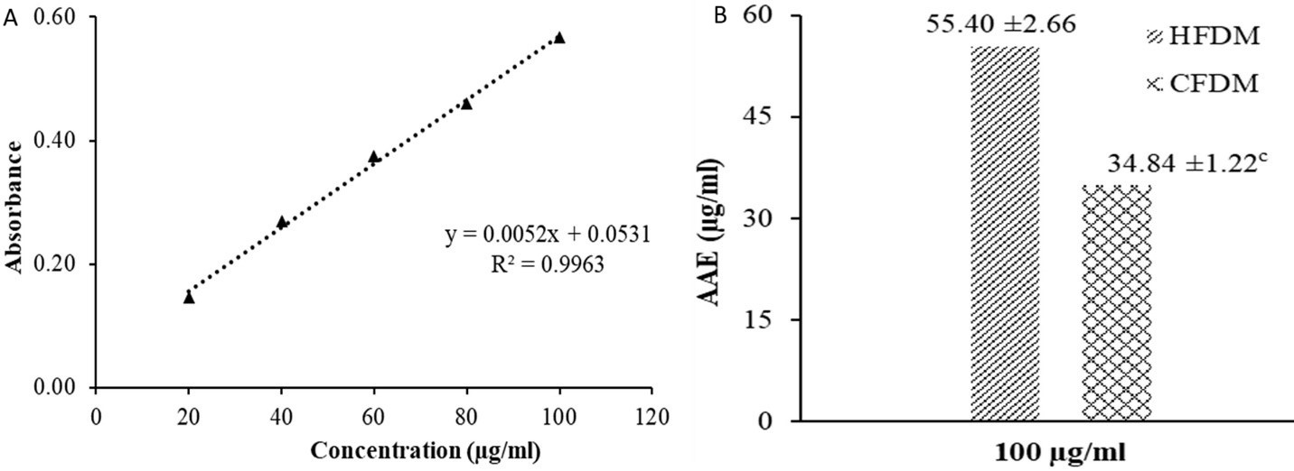
The TAC of D. microcarpum is depicted in Figure 1, revealing the AA calibration curve and the TAC of the HFDM and CFDM of D. microcarpum. The HFDM (55.40 ± 2.66 AAE µg/mL) showed a significantly (p<0.05) higher TAC than the CFDM (34.84 ± 1.22 AAE µg/mL) at the tested concentration (100 µg/mL).
Ferric Reducing Antioxidant Power
Figure 2 depicts the FRAP of D. microcarpum, showing the AA calibration curve and the FRAP of the HFDM and CFDM. The CFDM (57.84 ± 2.16 AAE µg/mL) exhibited a significantly (p<0.05) higher FRAP than the HFDM (46.11 ± 1.91 AAE µg/mL) at the tested concentration, contrary to the result for the TAC.
Ferric Thiocyanate Assay
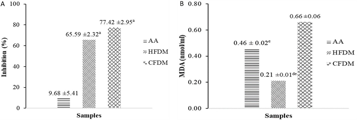
Figure 3 shows the anti-lipid peroxidation potential of D. microcarpum in the early stage of lipid peroxidation, revealing the percentage inhibitory effect of HFDM and CFDM. Although there was no significant (p>0.05) difference between the HFDM (65.59 ± 2.32%) and CFDM (77.42 ± 2.95%), both fractions exhibited significantly (p<0.05) higher percentage inhibition than AA (9.68 ± 5.41%).

Figure 1. TAC of D. microcarpum. (A) AA calibration curve and (B) TAC of the HFDM and CFDM. (c, p<0.05) is significantly lower than HFDM.

Figure 2. FRAP of D. microcarpum. (A) AA calibration curve and (B) FRAP of the HFDM and CFDM. (b, p<0.00) is significantly higher than HFDM.

Figure 3. Anti-lipid peroxidation potential of D. microcarpum. (A) FTC assay and (B) TBA assay. (a, p<0.05) is significantly higher than AA while (d and e, p<0.05) are significantly lower than AA and CFDM, respectively.
Thiobarbituric Acid Assay
The effect of D. microcarpum on the late stage of lipid peroxidation (MDA formation) is shown in Figure 3, showing the MDA concentrations. The HFDM (0.21 ± 0.01 nmol/mL) exhibited a significantly lower MDA concentration than the CFDM (0.66 ± 0.06 nmol/mL) and AA (0.46 ± 0.02 nmol/mL). Moreover, AA (0.46 ± 0.02 nmol/mL) showed a significantly lower MDA concentration than CFDM (0.66 ± 0.06 nmol/mL).
The present study explored the ability of D. microcarpum to scavenge free radicals and exert anti-lipid peroxidation effects in vitro via different techniques. The antioxidant activity of plants might be influenced by their phytochemical composition, including the ones identified in our study (69). The radical scavenging activity of the HFDM was superior to that of the CFDM in the phosphomolybdate assay, which might be attributed to the presence of a higher number of electron-donating compounds in the HFDM. Moreover, the HFDM was initially fractionated from the crude extract based on polarity, which might lead to more of these compounds in the HFDM than the CFDM. In contrast, the CFDM showed superior activity in the FRAP assay, which might be due to the preference of the compounds present in the CFDM for the ferric ions over the Mo (VI) ions (70). The FRAP and phosphomolybdate assays are principled in reducing ferric to ferrous ions and Mo (VI) to Mo (V), respectively. Thus, both methods determine the radical scavenging potential of a test sample. In the FTC and TBA assay, the anti-lipid peroxidation showed the superiority of the CFDM in the early stage of lipid peroxidation; however, the HFDM exhibited superior activity in the late stage of lipid peroxidation. This might be related to the phytochemical components present in these fractions, which in turn influence the anti-lipid peroxidation potential of the fractions at different stages.
Conclusion
The present study explored the phytoconstituents and free radical scavenging potential of D. microcarpum for its potential application in phytotherapy of oxidative stress and related ailments. Conclusively, the n-hexane and chloroform fractions of the plants showed promising free radical scavenging potential, which might be attributed to the phytochemicals detected in the plant with potential application in the phytotherapy of diseases linked to oxidative stress. Moreover, both fractions might serve as an invaluable source of novel compounds with antioxidant activity. Thus, further studies to identify and isolate these compounds are warranted.
Abbreviations
HFDM = n-hexane Fraction of D. microcarpum; CFDM = Chloroform Fraction of D. microcarpum; MDA = Malonaldehyde; ROS = Reactive Oxygen Species; RNS = Reactive Nitrogen species; TAC = Total Antioxidant Capacity; FRAP = Ferric Reducing Antioxidant Power; FTC = Ferric Thiocyanate; TBA = Thiobarbituric Acid; SEM = Standard Error of Mean
Declarations
Acknowledgment
The authors appreciate the contribution and institutional support of the Pharmaceutical Technology and Science Laboratory Technology Departments of Adamawa State Polytechnic, Yola.
Ethics Statement
Not applicable.
Data Availability
The unpublished data is available upon request to the corresponding author.
Funding Information
Not applicable.
Conflict of Interest
The authors declare no conflicting interest.
References
- Hunyadi A. The mechanism (s) of action of antioxidants: From scavenging reactive oxygen/nitrogen species to redox signaling and the generation of bioactive secondary metabolites. Med Res Rev. 2019;39(6):2505-33.
- Ali SS, Ahsan H, Zia MK, Siddiqui T, Khan FH. Understanding oxidants and antioxidants: Classical team with new players. J Food Biochem. 2020;44(3):e13145.
- Barreca D. Mechanisms of plant antioxidants action. Plants. 2020;10(1):35.
- Miftode A. Antioxidants: mechanisms of action and therapeutic applications. Medicro. 2022;6:1-8.
- Kıran TR, Otlu O, Karabulut AB. Oxidative stress and antioxidants in health and disease. Journal of Laboratory Medicine. 2023;47(1):1-11.
- Türkoğlu M, Pekmezci E, Sevinç H. Antioxidants and Antiaging. In: Alasalvar C, Shahidi F, Ho C-T, editors. Dietary Supplements with Antioxidant Activity: Understanding Mechanisms and Potential Health Benefits: The Royal Society of Chemistry; 2023. p. 0.
- Gęgotek A, Skrzydlewska E. Chapter Nine - Ascorbic acid as antioxidant. In: Litwack G, editor. Vitam Horm. 121: Academic Press; 2023. p. 247-70.
- Mathew BB, Tiwari A, Jatawa SK. Free radicals and antioxidants: A review. J Pharm Res. 2011;4(12):4340-3.
- Gholamian-Dehkordi N, Luther T, Asadi-Samani M, Mahmoudian-Sani MR. An overview on natural antioxidants for oxidative stress reduction in cancers; a systematic review. Immunopathol Persa. 2017;3(2):e12.
- Wang W, Kang PM. Oxidative stress and antioxidant treatments in cardiovascular diseases. Antioxidants. 2020;9(12):1292.
- Zhang P, Li T, Wu X, Nice EC, Huang C, Zhang Y. Oxidative stress and diabetes: antioxidative strategies. Front Med. 2020;14(5):583-600.
- Mozzini C, Pagani M. Oxidative stress in chronic and age-related diseases. Antioxidants. 2022;11(3):521.
- Ighodaro OM. Molecular pathways associated with oxidative stress in diabetes mellitus. Biomed Pharmacother. 2018;108:656-62.
- Klaunig JE. Oxidative Stress and Cancer. Curr Pharm Des. 2018;24(40):4771-8.
- Oguntibeju OO. Type 2 diabetes mellitus, oxidative stress and inflammation: examining the links. Int J Physiol Pathophysiol Pharmacol. 2019;11(3):45–63.
- Hayes JD, Dinkova-Kostova AT, Tew KD. Oxidative Stress in Cancer. Cancer Cell. 2020;38(2):167-97.
- Chikezie PC, Ojiako OA, Ogbuji AC. Oxidative stress in diabetes mellitus. Int J Biol Chem. 2015;9(3):92-109.
- Domingueti CP, Dusse LMSA, Carvalho MdG, de Sousa LP, Gomes KB, Fernandes AP. Diabetes mellitus: The linkage between oxidative stress, inflammation, hypercoagulability and vascular complications. J Diabetes Complications. 2016;30(4):738-45.
- Bhatti JS, Sehrawat A, Mishra J, Sidhu IS, Navik U, Khullar N, et al. Oxidative stress in the pathophysiology of type 2 diabetes and related complications: Current therapeutics strategies and future perspectives. Free Radical Biol Med. 2022;184:114-34.
- Dahiru MM, Badgal EB, Musa N. Phytochemistry, GS-MS analysis, and heavy metals composition of aqueous and ethanol stem bark extracts of Ximenia americana. GSC Biol Pharm Sci. 2022;21(3):145-56.
- Dahiru MM, Nadro MS. Anti-diabetic potential of Hyphaene thebaica fruit in streptozotocin-induced diabetic rats. J Expl and Molec Biol. 2022;23(1):29-36.
- Dahiru MM, Nadro SM. Hypolipidemic Potential of Ethyl acetate Extract of Hyphaene thebaica Fruit in Streptozotocin-induced Diabetic Rats. Majalah Obat Tradisional. 2022;27(2):159-64.
- Badgal EB, Dahiru MM, Musa N. Phytochemical profiling, heavy metals composition, in silico aphrodisiac potential, and ADMET study of Gardenia erubescens. Sciences of Phytochemistry. 2023;2(2):91-106.
- Dahiru MM, Abaka AM, Musa N. Phytochemical Analysis, In-vitro, and In-silico Antibacterial Activity of Stembark Extract of Anogeissus leiocarpus (DC) Guill and Perr. Sciences of Pharmacy. 2023;2(3):24-41.
- Dahiru MM, Badgal EB, Neksumi M. Phytochemical profiling and heavy metals composition of aqueous and ethanol extracts of Anogeissus leiocarpus. Journal of Faculty of Pharmacy of Ankara University. 2023;47(2):311-23.
- Dahiru MM, Musa N, Abaka AM, Abubakar MA. Potential Antidiabetic Compounds from Anogeissus leiocarpus: Molecular Docking, Molecular Dynamic Simulation, and ADMET Studies. Borneo J Pharm. 2023;6(3):249-77.
- Abaka AM, Dahiru MM, Abubakar KB, Luka J, Abubakar A, Abdullahi TB, et al. Phytochemical Profile and Antibacterial Activity of Nigella Sativa against Biofilm-producing Bacteria Uropathogens. Biology, Medicine, & Natural Product Chemistry. 2024;13(1):141-6.
- Musa N, Dahiru MM, Badgal EB. Characterization, In Silico Antimalarial, Anti-inflammatory, Antioxidant, and ADMET Assessment of Neonauclea excelsa Merr. Sciences of Pharmacy. 2024;3(2):92-107.
- Andrés Juan C, Pérez de Lastra JM, Plou Gasca FJ, Pérez-Lebeña E. The chemistry of Reactive oxygen Species (ROS) revisited: Outlining their role in biological macromolecules (DNA, lipids and proteins) and induced pathologies. 2021.
- Dahiru MM, Nadro MS. Phytochemical Composition and Antioxidant Potential of Hyphaene thebaica Fruit. Borneo J Pharm. 2022;5(4):325-33.
- Dahiru MM, Ahmadi H, Faruk MU, Aminu H, Hamman, Abreme GC. Phytochemical Analysis and Antioxidant Potential of Ethylacetate Extract of Tamarindus Indica (Tamarind) Leaves by Frap Assay. Journal of Fundamental and Applied Pharmaceutical Science. 2023;3(2):45-53.
- Dahiru MM, Alfa MB, Abubakar MA, Abdulllahi AP. Assessment of in silico antioxidant, anti-inflammatory, and antidiabetic activites of Ximenia americana L. Olacaceae. Advances in Medical, Pharmaceutical and Dental Research. 2024;4(1):1-13.
- Dahiru MM, Musa N. Phytochemical Profiling, Antioxidant, Antidiabetic, and ADMET Study of Diospyros mespiliformis Leaf, Hochst Ex A. Dc Ebenaceae. J Fac Pharm Ankara/Ankara Ecz Fak Derg. 2024;48(2):412-35.
- Dahiru MM, Umar AS, Muhammad M, Fari II, Musa ZY. Phytoconstituents, Fourier-Transform Infrared Characterization, and Antioxidant Potential of Ethyl Acetate Extract of Corchorus olitorius (Malvaceae). Sciences of Phytochemistry. 2024;3(1):1-10.
- Dahıru MM, Musa N. GC-MS Analysis, Antioxidant, Antidiabetic Activity, and ADMET Study of Diospyros mespiliformis Hochst. Ex A. DC. Ebenaceae Stembark. Hacettepe University Journal of the Faculty of Pharmacy. 2024;44(3):198-219.
- Dahiru MM. Recent advances in the therapeutic potential phytochemicals in managing diabetes. Journal of Clinical and Basic Research. 2023;7(1):13-20.
- Dahiru MM, Abaka AM, Ya'u I. Antibacterial Potential of Ximenia americana L. Olacaceae: Molecular Docking, Molecular Dynamics, and ADMET Prediction. Journal of Pharmacy. 2024;4(1):51-67.
- Dahiru MM, Musa N. Antidiabetic Activity of Diospyros mespiliformis on Alloxan-Induced Diabetic Rats. Journal of Fundamental and Applied Pharmaceutical Science. 2024;5(1):11-20.
- Oyeleke MB, Owoyele BV. Saponins and flavonoids from Bacopa floribunda plant extract exhibit antioxidant and anti-inflammatory effects on amyloid beta 1-42-induced Alzheimer's disease in BALB/c mice. J Ethnopharmacol. 2022;288:114997.
- Hassanpour SH, Doroudi A. Review of the antioxidant potential of flavonoids as a subgroup of polyphenols and partial substitute for synthetic antioxidants. Avicenna journal of phytomedicine. 2023;13(4):354.
- Okolo CE, Akah PA, Uzodinma SU. Antidiabetic activity of the root extract of Detarium microcarpum (Fabacaee) Guill and Perr. Phytopharmacology. 2012;3(1):12-8.
- Mariod AA. Wild fruits: Composition, nutritional value and products: Springer; 2019.
- Dembele N, Somboro AA, Badiaga M, Cisse S, Cisse M, Togola I, et al. Ethnobotanical survey and phytochemical screening of aqueous extracts from Detarium microcarpum Guill. Perr in Mali. Journal of Medicinal Plants Research. 2022;16(10):280-7.
- Dahiru M, Saheed KO, Adamu TM. Antibacterial and phytochemicals status of Detarium microcarpum (guill and perr) stem bark. Natural Resources for Human Health. 2022;2(4):385-90.
- Dahiru MM, Abaka AM, Artimas SP. Phytochemical Analysis and Antibacterial Activity of Methanol and Ethyl Acetate Extracts of Detarium microcarpum Guill. & Perr. Biology, Medicine, & Natural Product Chemistry. 2023;12(1):281-8.
- Sowemimo AA, Pendota C, Okoh B, Omotosho T, Idika N, Adekunle AA, et al. Chemical composition, antimicrobial activity, proximate analysis and mineral content of the seed of Detarium senegalense JF Gmelin. Afr J Biotechnol. 2011;10(48):9875-9.
- Dogara AM. Biological activity and chemical composition of Detarium microcarpum Guill. and Perr—a systematic review. Advances in Pharmacological and Pharmaceutical Sciences. 2022;2022(1):7219401.
- David J, Kolawole JA, Alemika TE, Agwom FM, Ajima U. Analgesic and anti-inflammatory activities of the leaf extracts of Detarium microcarpum Guill & Perr (Fabaceae). Asian Journal of Pharmacy and Pharmacology. 2020;6(5):348-55.
- Rouamba A, Nkono BLNY, Ouedraogo V, Duceac IA, Compaoré M, Ouédraogo M. Biocompatibility assessment and antiproliferative activity of detarium microcarpum Guill and Perr fruit pulp extracts. Int J Phytomedicine. 2021;13(2):6.
- Sanusi SB, Lawal SM, Usman A, Musa FM, Ardo B, Way TB. Phytochemical analysis and antibacterial activity of stem bark extracts of detarium microcarpum against bacteria causing gastrointestinal tract infections in humans. Dutse Journal of Pure and Applied Sciences. 2022;8(1b):82-9.
- Kurmi UG, Modu B, Patel P, Abubakar F, Umar RB, Balkore HA, et al. Phytochemical screening and in-vitro antioxidant activity of ethanolic leaves extract of detarium microcarpum. World Journal of Pharmaceutical Research. 2021;10(5):84.
- David J, Agwom FM, Ojerinde SO, Ajima U, Alemika TE. Comparative Antioxidant Activities of Leaf Extracts of Detarium microcarpum Guill & Perr (Fabaceae). Tropical Journal of Natural Product Research. 2022;6(11).
- Emohchonne JU, Ndidi US, Kashim AS, Sofiyullahi I, Binta S, Abdulmalik I, et al. Phytochemical Composition, Antioxidant and Antitrypanosomal Properties of Leaves and Stem Bark Extract of Detarium microcarpum: An in vitro study. Nigerian Journal of Biochemistry and Molecular Biology. 2022;37(3):158-65.
- Evans WC. Trease and Evans' pharmacognosy: Elsevier Health Sciences; 2009. 608 p.
- Prieto P, Pineda M, Aguilar M. Spectrophotometric quantitation of antioxidant capacity through the formation of a phosphomolybdenum complex: specific application to the determination of vitamin E. Anal Biochem. 1999;269(2):337-41.
- Oyaizu M. Studies on products of browning reaction antioxidative activities of products of browning reaction prepared from glucosamine. The Japanese journal of nutrition and dietetics. 1986;44(6):307-15.
- Kikuzaki H, Nakatani N. Antioxidant effects of some ginger constituents. J Food Sci. 1993;58(6):1407-10.
- Wakeel A, Jan SA, Ullah I, Shinwari ZK, Xu M. Solvent polarity mediates phytochemical yield and antioxidant capacity of Isatis tinctoria. PeerJ. 2019;7:e7857.
- Sharma S, Kumari A, Dhatwalia J, Guleria I, Lal S, Upadhyay N, et al. Effect of solvents extraction on phytochemical profile and biological activities of two Ocimum species: A comparative study. Journal of Applied Research on Medicinal and Aromatic Plants. 2021;25:100348.
- Muzahid AA, Sharmin S, Hossain MS, Ahamed KU, Ahmed N, Yeasmin MS, et al. Analysis of bioactive compounds present in different crude extracts of Benincasa hispida and Cucurbita moschata seeds by gas chromatography-mass spectrometry. Heliyon. 2023;9(1):e12702.
- Friday C, Akwada U, Igwe OU. Phytochemical screening and antimicrobial studies of Afzelia africana and Detarium microcarpum seeds. Chemistry International. 2018;4(3):170-6.
- Macáková K, Afonso R, Saso L, Mladěnka P. The influence of alkaloids on oxidative stress and related signaling pathways. Free Radical Biol Med. 2019;134:429-44.
- Uthman T, Salman R, Lamorde I. Free Radical Scavenging Activity of Alkaloid and Flavonoid Fractions of Kalanchoe pinnata Leaves. Journal of Drug Design and Medicinal Chemistry. 2023.
- Dalimunthe A, Hasibuan PAZ, Silalahi J, Sinaga SF, Satria D. Antioxidant activity of alkaloid compounds from Litsea cubeba Lour. Oriental Journal of Chemistry. 2018;34(2):1149.
- Lokesh R, Manasvi V, Lakshmi BP. Antibacterial and antioxidant activity of saponin from Abutilon indicum leaves. Asian J Pharm Clin Res. 2016;9(3):344-7.
- Odeyemi S, Afolayan A, Bradley G. In vitro anti-inflammatory and free radical scavenging activities of crude saponins extracted from Albuca bracteataJacq. Bulb. African Journal of Traditional, Complementary and Alternative Medicines. 2015;12(4):34-40.
- Xie J-H, Dong C-j, Nie S-P, Li F, Wang Z-J, Shen M-Y, et al. Extraction, chemical composition and antioxidant activity of flavonoids from Cyclocarya paliurus (Batal.) Iljinskaja leaves. Food Chem. 2015;186:97-105.
- Badmus JA, Ekpo OE, Rautenbach F, Marnewick JL, Hussein AA, Hiss DC. Isolation and antioxidant activity of flavonoids from Holarrhena floribunda (G. don) leaves. Acta Biochim Pol. 2016;63(2):353-8.
- Farag RS, Abdel-Latif MS, Abd El Baky HH, Tawfeek LS. Phytochemical screening and antioxidant activity of some medicinal plants’ crude juices. Biotechnology Reports. 2020;28:e00536.
- Christodoulou MC, Orellana Palacios JC, Hesami G, Jafarzadeh S, Lorenzo JM, Domínguez R, et al. Spectrophotometric methods for measurement of antioxidant activity in food and pharmaceuticals. Antioxidants. 2022;11(11):2213.

 ETFLIN
Notification


