
RESEARCH ARTICLE
Nano Liposomal Curcumin as an Adjuvant: Enhancing Cisplatin Anticancer Effects in HeLa Cells
Academic Editor:
Sciences of Pharmacy|Vol. 4, Issue 2, pp. 96-102 (2025)
CC BY 4.0-2025 Authors
Views
Downloads
Shares
Received
May 10, 2025Revised
Jun 4, 2025Accepted
Jun 7, 2025Published
Jun 16, 2025
Abstract
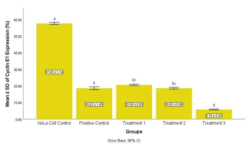
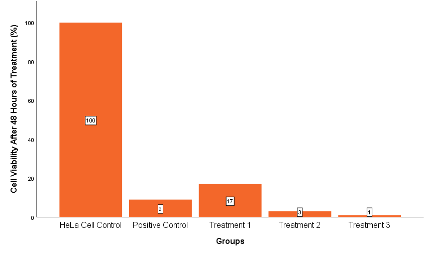
Cervical cancer, mainly driven by oncogenic HPV infections, remains a global health burden. Cisplatin is standard chemotherapy for advanced cases but is limited by toxicity. Nano liposomal curcumin, with improved bioavailability, may enhance cisplatin’s efficacy. This study investigated the combination’s effect on HeLa cells by analyzing Cyclin E1 and Bcl-2 expression. Nano liposomal curcumin was synthesized using thin-film hydration, yielding stable 32 nm nanoparticles. HeLa cells were divided into control and treatment groups, and varying doses of nano liposomal curcumin with cisplatin were received. Flow cytometry revealed significant reductions in Cyclin E1 (from 18.62 ± 1.45 to 5.79 ± 0.47) and Bcl-2 (from 32.56 ± 0.82 to 28.19 ± 0.30) at the highest dose (p < 0.05). Cell viability decreased to 9% with cisplatin alone and 1% with the combination. These results indicate that nano liposomal curcumin enhances cisplatin’s antiproliferative and pro-apoptotic effects, supporting its potential as an adjuvant to lower cisplatin doses while maintaining efficacy. Further research involving additional molecular markers, in vivo models, and clinical trials is needed to optimize dosing, confirm safety, and evaluate therapeutic potential.
Introduction
Globally, cervical cancer is the fourth most common cancer, with 660,000 new cases and 350, 000 deaths in 2022 (1). In Indonesia, the estimated number of cases reached 408,661, resulting in 242, 988 deaths (2). Most cases are associated with high-risk human papillomavirus (HPV), particularly types 16 and 18, which play a central role in cervical carcinogenesis (3).
These oncogenic HPV types disrupt the cell cycle as E6 and E7 oncoproteins, which inactivate p53 and pRb, leading to the upregulation of Cyclin E1 and increased expression of the anti-apoptotic protein B-cell lymphoma 2 (Bcl-2), thereby promoting cell proliferation and resistance to apoptosis (4-6). Cisplatin remains the primary chemotherapy for advanced cervical cancer (7). Cisplatin inhibits proliferation and induces apoptosis through p53 activation while downregulating Cyclin E1 and Bcl-2 expression (8). However, its effectiveness is often limited by toxic side effects on healthy cells (9). Therefore, adjuvant strategies are needed to enhance cisplatin efficacy while minimizing its side effects (10).
Curcumin, a bioactive compound in Curcuma longa L., exerts anticancer effects by increasing the production of reactive oxygen species (ROS), activating p53, upregulating p21, and ultimately downregulating Cyclin E1 and Bcl-2 expression (11-15). The combination treatment of curcumin and cisplatin has been shown to improve chemotherapy sensitivity by reducing proliferation and enhancing apoptosis, yet curcumin’s low bioavailability limits its therapeutic application (10, 16, 17). Nanotechnology-based formulations, collectively termed nanocurcumin, have been developed to overcome these limitations and improve curcumin’s therapeutic effects on cancer cells. Among the formulations, nano liposomal curcumin encapsulates curcumin within liposomal vesicles, substantially improving its stability, solubility, and bioavailability, facilitating enhanced delivery to cancer cells (10, 18). Previous studies indicate that nanocurcumin can enhance the effects of cisplatin (19, 20).
Despite the known individual effects of nano liposomal curcumin and cisplatin, their combined impact on cervical cancer cells remains unexplored. This study investigates the synergistic potential of nano liposomal curcumin and cisplatin in HeLa cells by analyzing Cyclin E1 and Bcl-2 expression, focusing on proliferation inhibition and apoptosis induction. We hypothesize that the combination treatment will synergistically inhibit proliferation and induce apoptosis in HeLa cells by downregulating Cyclin E1 and the anti-apoptotic protein Bcl-2 expression.
Material and Methodology
Research Ethics
This study adhered to ethical guidelines and received approval from the Health Research Ethics Committee, Faculty of Medicine, Universitas Brawijaya. Ethical clearance was granted through an exemption letter No. 488/EC/KEPK-S2/12/2024 for the analysis of Cyclin E1 expression and No. 02/EC/KEPK-S2/01/2024 for the analysis of Bcl-2 expression. The research protocol was reviewed and declared exempt from full ethical review.
Materials
The synthesis of nano liposomal curcumin was carried out using turmeric rhizome (Materia Medika, Indonesia), ethanol (96%, Merck, Germany), isopropyl alcohol (Merck, Germany), Tween 80 (Sigma-Aldrich, USA), soy phosphatidylcholine (Lipoid GmbH, Germany), cholesterol (Sigma-Aldrich, USA), chloroform (Merck, Germany), and PBS (Bioworld, USA). The preparation process employed a Soxhlet extractor (Buchi, Switzerland), rotary evaporator (Heidolph, Germany), probe sonicator (Qsonica Q700, USA), magnetic stirrer (Stuart CB162, Stuart Equipment Ltd., UK), particle size analyzer (Shimadzu SALD-7500nano, Shimadzu Corporation, Japan), UV-Vis spectrophotometer (Jenway 6800, Cole-Parmer, UK), and shaker incubator (Stuart SI500, Stuart Equipment Ltd., UK).
HeLa cells, a human cervical cancer cell line, were obtained from the American Type Culture Collection (ATCC) through the cell collection of the Biomedical Laboratory, Faculty of Medicine, Universitas Brawijaya. Cells were maintained in RPMI-1640 medium (Gibco, Thermo Fisher Scientific, USA) supplemented with 10% fetal bovine serum (Biowest, France) and 1% antibiotic-antimycotic solution (Gibco, Thermo Fisher Scientific, USA) to prevent contamination. Trypsin-EDTA (Gibco, Thermo Fisher Scientific, USA) was used for cell detachment during harvesting. Cisplatin (Kalbe, Indonesia) was used as the chemotherapeutic agent in the experiment. Cell culture materials included T-25 culture flasks (Biologix, China), 6-well plates (SPL, South Korea), centrifuge tubes (1.5 mL, 15 mL, and 50 mL; Biologix, China), centrifuge microtubes 1.5 mL, a centrifuge (Biosan, Latvia), a Class II biosafety cabinet (Safe Fast Elite, Safe Fast, Italy), a CO₂ incubator (ESCO, Singapore), and an inverted microscope (Olympus, Japan).
Flow cytometric analysis of Cyclin E1 expression was performed using centrifuge tubes (1.5 mL, 15 mL, and 50 mL; Biologix, China), round-bottom tubes, a vortex mixer (Thermo Scientific, USA), a centrifuge (Heraeus Sepatech, Germany), BD FACSCalibur flow cytometer (BD Biosciences, USA), and CellQuest Pro software (version 5.1, BD Biosciences, USA). The reagents included a PE-conjugated Cyclin E1 polyclonal antibody (Bioss, China, cat. bs-0573R-PE), cell staining buffer (BioLegend, China), PBS (Bioworld, USA), and Nuclear Factor Fixation and Permeabilization Buffers (BioLegend, China). For Bcl-2 expression analysis, the same instrumentation was used, along with a FITC-conjugated Bcl-2 (Ser87) polyclonal antibody (Bioss, China, cat. bs-12577R-FITC), Cyto-Fast™ Fix/Perm and Perm Wash Buffer Set (BioLegend, China), and deionized water (Widatra Bhakti, Indonesia).
Nano Liposomal Synthesis
Turmeric rhizomes were thoroughly cleaned, boiled, and sun-dried to a final % moisture content of 10%, then peeled and ground into a fine powder. A total of 15 g of turmeric powder was extracted using 96% ethanol in a Soxhlet apparatus at 60 °C for 8 h, yielding crude curcumin. From this extract, 1 g was purified by mixing with 25 mL of hexane, then drying and recrystallizing 10 mL of isopropyl alcohol at a 1: 1.53 molar ratio to obtain pure curcumin crystals.
Nano liposomal curcumin was synthesized using the thin-film hydration method based on the formulation described by Chen et al. (2012). Briefly, 2 mg of curcumin, 40 mg of soy phosphatidylcholine (SPC), 5 mg of cholesterol, and 5 mg of Tween-80 were dissolved in chloroform. The solvent was evaporated using a rotary evaporator at 50 °C for 60 min to form a thin lipid film. The lipid layer was then hydrated with 1 mL of phosphate-buffered saline (PBS, pH 6.5) containing Tween-80 at 60 °C for 30 min. The resulting liposomal suspension was sonicated at 80 W for 3 min to produce uniform nanoparticles (21). Particle size was analyzed using a Particle Size Analyzer (PSA), and curcumin was identified via High-Performance Liquid Chromatography (HPLC).
Cell Culture and Treatments
HeLa cells (passage number 45) were thawed at 37 °C and cultured in RPMI-1640 complete medium (CM) supplemented with 10% FBS and 1% antibiotic-antimycotic. After thawing, cells were seeded into a T25 flask containing 5 mL CM and incubated at 37 °C in a humidified 5% CO₂ atmosphere for 3 days to allow recovery and proliferation, with one medium change performed after 48 h. Following this incubation period, cells were harvested using Trypsin-EDTA, and cell viability was assessed using the trypan blue exclusion assay.
After confirming cell viability, HeLa cells were counted using a hemocytometer, and 5 × 10⁵ viable cells were seeded into each of 15 wells across three 6-well plates, corresponding to five experimental groups with three replicates each. After overnight incubation, cells were treated according to group designation: (1) HeLa cell control (CM only), (2) positive control (cisplatin 5 µg/mL), and (3-5) combination treatments of cisplatin (2.5 µg/mL) with nano liposomal curcumin at 25, 50, or 100 µg/mL.
The cisplatin concentrations were selected based on Li et al. (2021), who reported that 5 µg/mL cisplatin inhibited cervical cancer cell proliferation by 17.43% (22). This dose is below the reported LD₅₀ of cisplatin (approximately 10.5 µg/mL), minimizing non-specific cell death caused by excessive toxicity (23). To evaluate whether nano liposomal curcumin could enhance cisplatin’s therapeutic efficacy while reducing its cytotoxicity, a lower dose of 2.5 µg/mL was used for the combination treatments. This intentional dose reduction aimed to investigate potential synergistic effects with nano liposomal curcumin and to explore the feasibility of minimizing cisplatin-related toxicity, a common goal in combination therapy.
Nano liposomal curcumin concentrations of 25, 50, and 100 µg/mL were selected based on preliminary CCK-8 assays, which indicated effective inhibition of HeLa cell proliferation within 48 h post-treatment. The results of this viability test are presented in the Results section and were used to guide dose selection for the flow cytometry experiment. These doses were chosen considering their safety profiles. In vivo data indicate that curcumin has very low systemic toxicity, with an oral LD₅₀ value of > 5000 mg/kg body weight in rats (24), supporting the rationale for selecting nano liposomal curcumin doses that are both effective and safe.
After 48 h, cells cultured in 6-well plates were harvested using trypsin-EDTA, incubated for 3 min at 37 °C until detachment, followed by centrifugation at 500 × g for 5 min using a centrifuge (Biosan, Latvia). Cell pellets were then washed with PBS and prepared for flow cytometry analysis.
Flow Cytometry Analysis
Harvested cells were fixed using Nuclear Factor Fixation Buffer (for Cyclin E1) and Cyto-Fast™ Fix/Perm Buffer (for Bcl-2). Permeabilization was performed with Nuclear Factor Permeabilization Buffer and Cyto-Fast™ Perm Wash Solution. Cells were incubated with PE-conjugated Cyclin E1 Polyclonal Antibody (bs-0573R-PE) for 30 min to allow detection, and FITC-conjugated Bcl-2 (Ser87) Polyclonal Antibody (bs-12577R-FITC) in the dark at room temperature for 20 min. Following staining, cells were rinsed, suspended in 300 µL buffer, and analyzed using a BD FACSCalibur flow cytometer (BD Biosciences, USA). Data were processed with CellQuest Pro software (version 5.1, BD Biosciences, USA) to quantify fluorescence intensity, indicating Cyclin E1 and Bcl-2 expression.
| Identity | Retention Time | Area | Height | Theoretical Plate | Tailing Factor |
|---|---|---|---|---|---|
| Nano Liposomal Curcumin Sample | 9.504 | 616.531 | 27.442 | 4865.895 | 1.122 |
| Curcumin Standard | 9.484 | 680.523 | 28.795 | 4552.908 | 1.114 |
Statistical Analysis
Data analysis was performed using IBM SPSS Statistics 26. A one-way ANOVA was applied, followed by Tukey’s HSD test for post-hoc analysis. Statistical significance was defined as p < 0.05.
Result and Discussion
Nano Liposomal Curcumin Characterization
Characterization of Nano Liposomal Curcumin
Particle size analysis (PSA) showed that the nano liposomal curcumin had an average diameter of 32 nm, placing them well within the nanoscale range (20–150 nm) (25). The Polydispersity Index (PDI) was 0.1633, indicating a uniform particle distribution. The zeta potential was −10.69 mV, suggesting moderate colloidal stability. The absorbance of the formulation was 0.869 ± 0.093, indicating consistent concentration across replicates. These characteristics suggest that the liposomal formulation may enhance curcumin's stability, cellular uptake, and therapeutic delivery.
Curcumin Identification and Purity
High-performance liquid chromatography (HPLC) analysis was conducted to confirm nano liposomal curcumin's identity, purity, and stability. The retention time of the nano liposomal curcumin sample was 9.504 min, closely matching that of the curcumin standard (9.484 min, Merck Catalog No. 8.20354.0010). The peak area of the sample was 616.531, and the peak height was 27.442, compared to 680.523 and 28.795, respectively, for the standard. These results demonstrate that the sample contains curcumin as the major component with high purity, based on the overlapping retention time and consistent chromatographic profile. According to Kurniawan (2012), such similarity in retention time and peak characteristics is a valid indicator of both compound identity and purity (26).
Theoretical plate number (4865.895) and tailing factor (1.122) further support the validity and sharpness of the peak, indicating optimal separation performance and confirming the purity and homogeneity of the sample. These values fulfill the criteria proposed by Chulikhit et al. (2023), which state that a theoretical plate number > 2000 and tailing factor ≤2 reflect efficient chromatographic separation and accurate compound identification (27).
Effects on Cyclin E1 Expression
Cyclin E1 is a key regulator of the G1/S phase transition and is a well-established cellular proliferation marker. Its overexpression accelerates entry into the S phase, increasing the risk of uncontrolled cell proliferation. In cervical cancer, elevated Cyclin E1 expression is primarily driven by the activity of human papillomavirus (HPV) oncoproteins E6 and E7, which disrupt normal cell cycle regulation (28, 5). As a first-line chemotherapeutic agent, cisplatin inhibits Cyclin E1 expression by inducing DNA damage, which subsequently activates p53. This activation promotes the transcription of p21, a cyclin-dependent kinase inhibitor that suppresses Cyclin E/CDK2 activity, resulting in cell cycle arrest (29, 9).
This study used flow cytometry to evaluate the overall expression of Cyclin E1 following treatment. The results revealed that cisplatin at 5 µg/mL significantly reduced Cyclin E1 expression compared to the control HeLa cells. More notably, combining cisplatin (2.5 µg/mL) with nano liposomal curcumin further suppressed Cyclin E1 expression in a dose-dependent manner. The most significant reduction was observed in Treatment Group 3 (cisplatin 2.5 µg/mL + nano liposomal curcumin 100 µg/mL), with a statistically significant difference compared to all other groups (p < 0.05). However, no significant suppression was observed in Treatment Groups 1 and 2, which received lower nano liposomal curcumin concentrations (25 and 50 µg/mL), indicating that optimal inhibitory effects require higher doses. Differences in Cyclin E1 expression among groups can be seen in Figure 1.
The effectiveness of nanocurcumin in downregulating Cyclin E1 has also been demonstrated in other cancer types. Li et al. (2022) reported that curcumin inhibited colon cancer cell proliferation by reducing Cyclin E1 expression through p21 upregulation (14). As a CDK inhibitor, p21 downregulates Cyclin E1/CDK2 activity, thereby delaying the G1/S transition. Using nanocurcumin improves curcumin's cellular uptake and bioavailability, enhancing its pharmacological efficacy (8, 17).
The underlying mechanism for this synergistic effect involves inhibiting the NF-κB signaling pathway. Nanocurcumin directly interacts with IKKβ, preventing IκB phosphorylation and subsequent NF-κB activation. This inhibition promotes p53 activation and upregulates p21 expression, leading to suppressed Cyclin E1 levels (30, 31). Furthermore, nanocurcumin has proven to stabilize structurally mutated p53 proteins, such as p53Y220C, restoring their tumor suppressor function (14, 32).
These findings suggest that the combination of cisplatin and nano liposomal curcumin, particularly at a dose of 2.5 µg/mL cisplatin and 100 µg/mL nano liposomal curcumin, significantly downregulates Cyclin E1 expression. This indicates that nano liposomal curcumin may serve as a potent adjuvant to enhance cisplatin’s efficacy in cervical cancer therapy by targeting key regulators of cell proliferation. However, further investigations using additional molecular markers are necessary to validate these findings and fully elucidate the underlying mechanisms.
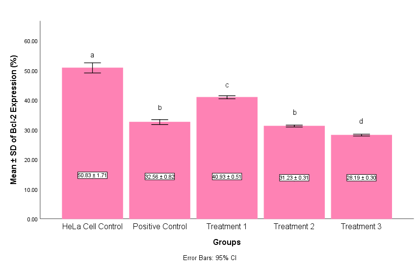
Effects on Bcl-2 Expression
Our results showed a significant reduction in Bcl-2 expression in HeLa cells following treatment with nano liposomal curcumin and cisplatin, supporting the hypothesis that this combination effectively induces apoptosis by targeting anti-apoptotic mechanisms. The results showed that all treatment groups (positive control, treatment 1, treatment 2, and treatment 3) were significantly different from the untreated HeLa control group (p < 0.05). However, Bcl-2 expression in the positive control group was not significantly different from treatment 2, indicating that this dose combination had a comparable effect to cisplatin monotherapy in reducing Bcl-2 expression. Notably, the highest nano liposomal curcumin dose combined with cisplatin (treatment 3) produced the most pronounced decrease in Bcl-2, even outperforming cisplatin monotherapy. This suggests a synergistic effect of nano liposomal curcumin in enhancing cisplatin’s pro-apoptotic activity.
These findings align with the established role of Bcl-2 as a key anti-apoptotic protein that preserves mitochondrial integrity and inhibits cell death, often upregulated in cancer cells to evade apoptosis (33, 34). The observed downregulation of Bcl-2 may result from activation of p53, a tumor suppressor protein known to repress Bcl-2 expression (35, 9). Cisplatin induces DNA damage that activates p53, while nano liposomal curcumin appears to amplify this effect by increasing intracellular reactive oxygen species (ROS), further promoting p53 activation and apoptotic signaling (36, 6). Supporting literature corroborates this mechanism. Gupta et al. (2020) reported that nanocurcumin increases ROS levels in cancer cells, triggering oxidative stress and apoptosis (37). Zhang et al. (2023) similarly showed that elevated ROS reduces Bcl-2 expression in HeLa cells, linking oxidative stress to intrinsic apoptotic pathway activation (38). This involves mitochondrial dysfunction and subsequent caspase activation, which further suppresses Bcl-2 and facilitates programmed cell death.
Moreover, studies by Moawad et al. (2023), Wang et al. (2020), and Shang et al. (2016) emphasize the capacity of curcumin formulations to upregulate p53, reinforcing the downregulation of Bcl-2 and potentiation of apoptosis (39, 15, 11). Our data extend these findings by demonstrating that nano liposomal curcumin combined with cisplatin effectively modulates these pathways, supporting its potential as an adjuvant therapy in cervical cancer.
Although no post-treatment viability test was performed in the main experiment, dose selection was guided by preliminary viability data (Figure 3), which demonstrated that the chosen concentrations of nano liposomal curcumin and cisplatin were sufficient to significantly reduce HeLa cell proliferation. These findings support the plausibility of apoptosis induction through reduced Bcl-2 expression in the treatment groups.
In the present study, it is hypothesized that nano liposomal curcumin enhanced the cisplatin-induced activation of p53, leading to greater suppression of Bcl-2 expression and a more robust activation of the apoptotic pathway. These results suggest a potential synergistic effect in activating the intrinsic apoptotic pathway. Further studies are needed to explore the underlying molecular mechanisms in greater detail, including the expression of other apoptosis-related proteins. Expanding the dose range of nano liposomal curcumin and cisplatin is also recommended to identify the optimal therapeutic window. Moreover, in vivo and clinical studies are warranted to evaluate the safety and efficacy of this combination in more complex biological systems.
Conclusion
These findings highlight nano liposomal curcumin’s potential as a cisplatin adjuvant by inhibiting cell proliferation through Cyclin E1 suppression and promoting apoptosis via Bcl-2 downregulation. Given its ability to enhance cisplatin’s anticancer effect at lower doses, nano liposomal curcumin may serve as a promising adjunct in cervical cancer therapy to improve treatment outcomes and reduce chemotherapy-related side effects. Nevertheless, further biomolecular studies are necessary to confirm these mechanisms and support clinical translation, including assessment of other apoptosis-related proteins, expanded dose ranges, and in vivo and clinical validation.
Abbreviations
HPV = Human Papillomavirus; pRb = Protein Retinoblastoma; Bcl-2 = B-Cell Lymphoma 2; ROS = Reactive Oxygen Species; PSA = Particle Size Analyzer; HPLC = High-Performance Liquid Chromatography; CM = Complete Medium; PE = Phycoerythrin; FITC = Fluorescein Isothiocyanate; DNA = Deoxyribonucleic Acid; CDK = Cyclin Dependent Kinase; NF-κB = Nuclear Factor Kappa B; IKK = IkB kinase; IκB = Inhibitor of Kappa B; CGM = Curcumin-Galactomannoside Complex; CNCs = Curcumin Nanocapsules.
Declarations
Acknowledgment
We thank Saiful Arifin and Wahyudha Ngatiril Lady for their laboratory support, and the Biomedical Laboratory, Faculty of Medicine, Universitas Brawijaya, for facilitating this research.
Conflict of Interest
The authors declare no conflict of interest.
Data Availability
The data supporting the findings of this study are available from the corresponding author upon reasonable request.
Ethics Statement
This study complied with ethical standards and received approval from the Health Research Ethics Committee, Faculty of Medicine, Universitas Brawijaya. Ethical clearance was provided via exemption letters No. 488/EC/KEPK-S2/12/2024 for Cyclin E1 analysis and No. 02/EC/KEPK-S2/01/2024 for Bcl-2 analysis. The research protocol was reviewed and deemed exempt from full ethical review.
Funding Information
The author(s) declare that no financial support was received for the research, authorship, and/or publication of this article.
References
- World Health Organization. Cervical cancer [Internet]. 2024 [cited 2024 Sep 5]. Available from: https://www.who.int/news-room/fact-sheets/detail/cervical-cancer.
- Ministry of Health RI. Ministry of Health determined to accelerate cervical cancer elimination [Internet]. 2024 [cited 2024 Sep 5]. Available from: https://sehatnegeriku.kemkes.go.id/baca/rilis-media/20240222/4144973/kemenkes-bertekad-mempercepat-eliminasi-kanker-serviks/.
- Zhang C, Zhu Q, Gu J, Chen S, Li Q, Ying L. Down-regulation of CCNE1 expression suppresses cell proliferation and sensitizes gastric carcinoma cells to cisplatin. Biosci Rep. 2020;39(6):BSR20190381.
- Pal A, Kundu R. Human papillomavirus E6 and E7: the cervical cancer hallmarks and targets for therapy. Front Microbiol. 2020;10:3116.
- Tavakolian S, Goudarzi H, Faghihloo E. Cyclin-dependent kinases and CDK inhibitors in virus-associated cancers. Infect Agents Cancer. 2020;15(1):27.
- Zheng Y, Zhang W, Xu L, Zhou H, Yuan M, Xu H. Recent progress in understanding the action of natural compounds at novel therapeutic drug targets for the treatment of liver cancer. Front Oncol. 2022;26(11):795548.
- Colombo N, Dubot C, Lorusso D, Caceres MV, Hasegawa K, Shapira-Frommer R, Monk BJ. Pembrolizumab for persistent, recurrent, or metastatic cervical cancer. N Engl J Med. 2021;385(20):1856-67.
- Dasari S, Njiki S, Mbemi A, Yedjou CG, Tchounwou PB. Pharmacological effects of cisplatin combination with natural products in cancer chemotherapy. Int J Mol Sci. 2022;23(3):1532.
- Tchounwou PB, Dasari S, Noubissi FK, Ray P, Kumar S. Advances in our understanding of the molecular mechanisms of action of cisplatin in cancer therapy. J Exp Pharmacol. 2021;18(13):303-28.
- Hussain Y, Islam L, Khan H, Filosa R, Aschner M, Javed S. Curcumin–cisplatin chemotherapy: a novel strategy in promoting chemotherapy efficacy and reducing side effects. Phytother Res. 2021;35(12):6514-25.
- Shang HS, Chang CH, Chou YR, Yeh MY, Au MK, Lu HF, Chung JG. Curcumin causes DNA damage and affects associated protein expression in HeLa human cervical cancer cells. Oncol Rep. 2016;36(4):2207-15.
- Gupta R, Jha A, Ambasta RK, Kumar P. Regulatory mechanism of cyclins and cyclin-dependent kinases in post-mitotic neuronal cell division. Life Sci. 2021;285:120006.
- Edwards RL, Luis PB, Nakashima F, Kunihiro AG, Presley SH, Funk JL, Schneider C. Mechanistic differences in the inhibition of NF-κB by turmeric and its curcuminoid constituents. J Agric Food Chem. 2020;68(22):6154-60.
- Li P, Pu S, Lin C, He L, Zhao H, Yang C, Zhou Z. Curcumin selectively induces colon cancer cell apoptosis and S cell cycle arrest by regulating Rb/E2F/p53 pathway. J Mol Struct. 2022;1263:133180.
- Wang T, Wu X, Al Rudaisat M, Song Y, Cheng H. Curcumin induces G2/M arrest and triggers autophagy, ROS generation and cell senescence in cervical cancer cells. J Cancer. 2020;11(22):6704-15.
- Park BH, Lim JE, Jeon HG, Seo SI, Lee HM, Choi HY, et al. Curcumin potentiates antitumor activity of cisplatin in bladder cancer cell lines via ROS-mediated activation of ERK1/2. Oncotarget. 2016;7(39):63870-86.
- Sandhiutami NMD, Arozal W, Louisa M, Rahmat D, Wuyung PE. Curcumin nanoparticle enhances the anticancer effect of cisplatin by inhibiting PI3K/AKT and JAK/STAT3 pathway in rat ovarian carcinoma induced by DMBA. Front Pharmacol. 2021;11:603235.
- Jacob S, Kather FS, Morsy MA, Boddu SH, Attimarad M, Shah J, Nair AB. Advances in nanocarrier systems for overcoming formulation challenges of curcumin: current insights. Nanomaterials (Basel). 2024;14(8):672.
- Mundekkad D, Cho WC. Applications of curcumin and its nanoforms in the treatment of cancer. Pharmaceutics. 2023;15(9):2223.
- Goswami S, Saxena S, Yadav S, Goswami D, Brahmachari K, Karmakar S, Brahmachari S. Review of curcumin and its different formulations: pharmacokinetics, pharmacodynamics and pharmacokinetic-pharmacodynamic interactions. OBM Integr Complement Med. 2022;7(4):1-35.
- Chen Y, Wu Q, Zhang Z, Yuan L, Liu X, Zhou L. Preparation of curcumin-loaded liposomes and evaluation of their skin permeation and pharmacodynamics. Molecules. 2012;17(5):5972–87.
- Li X, Zhao J, Yan T, Mu J, Lin Y, Chen J, Meng X. Cyanidin‐3‐O‐glucoside and cisplatin inhibit proliferation and downregulate the PI3K/AKT/mTOR pathway in cervical cancer cells. J Food Sci. 2021;86(6):2700-12.
- Rasoulian B, Kaeidi A, Rezaei M, Hajializadeh Z. Cellular preoxygenation partially attenuates the antitumoral effect of cisplatin despite highly protective effects on renal epithelial cells. Oxid Med Cell Longev. 2017;2017:720375.
- Aggarwal ML, Chacko KM, Kuruvilla BT. Systematic and comprehensive investigation of the toxicity of curcuminoid-essential oil complex: A bioavailable turmeric formulation. Mol Med Rep. 2015;13(1):592–604.
- Aguilar-Pérez KM, Medina DI, Parra-Saldívar R, Iqbal HMN. Nano-size characterization and antifungal evaluation of essential oil molecules-loaded nanoliposomes. Molecules. 2022;27(17):5728.
- Kurniawan FC. Determination of curcumin in capsule containing Curcuma xanthorrhiza Roxb. extracts by high performance liquid chromatography. Farmasains J Farmasi dan Ilmu Kesehatan. 2012;2(1).
- Chulikhit Y, Maneenet J, Monthakantirat O, Khamphukdee C, Chotritthirong Y, Limsakul S, Boonyarat C, Daodee S. Factors influencing high-performance liquid chromatography for piperine determination in traditional Thai formulas. J Appl Pharm Sci. 2023;13(9):48–57.
- Hemmat N, Baghi HB. The interaction of human papillomaviruses and adeno-associated viruses in suppressive co-infections. Infect Genet Evol. 2019;73:66-70.
- Tang Q, Wang X, Jin H, Mi Y, Liu L, Dong M, Zou Z. Cisplatin-induced ototoxicity: updates on molecular mechanisms and otoprotective strategies. Eur J Pharm Biopharm. 2021;163:60-71.
- Bortel N, Armeanu-Ebinger S, Schmid E, Kirchner B, Frank J, Kocher A, Ellerkamp V. Effects of curcumin in pediatric epithelial liver tumors: inhibition of tumor growth and alpha-fetoprotein in vitro and in vivo involving the NF-κB- and β-catenin pathways. Oncotarget. 2015;6(38):40680.
- Liang B, Liu Z, Cao Y, Zhu C, Zuo Y, Huang L, Bu X. MC37, a new mono-carbonyl curcumin analog, induces G2/M cell cycle arrest and mitochondria-mediated apoptosis in human colorectal cancer cells. Eur J Pharmacol. 2017;796:139-48.
- Wang W, Li M, Wang L, Chen L, Goh BC. Curcumin in cancer therapy: exploring molecular mechanisms and overcoming clinical challenges. Cancer Lett. 2023;570:216332.
- Carneiro BA, El-Deiry WS. Targeting apoptosis in cancer therapy. Nat Rev Clin Oncol. 2020;17(7):395-417.
- Saddam M, Paul SK, Habib MA. Emerging biomarkers and potential therapeutics of the BCL-2 protein family: the apoptotic and anti-apoptotic context. Egypt J Med Hum Genet. 2024;25(12).
- Chen Y, Wang C, Qi M, Wei Y, Jiang H, Du Z. Molecular targets of cisplatin in HeLa cells explored through competitive activity-based protein profiling strategy. J Inorg Biochem. 2024;254:112518.
- Abadi AJ, Mirzaei S, Mahabady MK, Hashemi F, Zabolian A, Hashemi F, Raee P, Aghamiri S, Ashrafizadeh M, Aref AR, Hamblin MR, Hushmandi K, Zarrabi A, Sethi G. Curcumin and its derivatives in cancer therapy: Potentiating antitumor activity of cisplatin and reducing side effects. Phytother Res. 2022;36(1):189-213.
- Gupta N, Verma K, Nalla S, Kulshreshtha A, Lall R, Prasad S. Free radicals as a double-edged sword: the cancer preventive and therapeutic roles of curcumin. Molecules. 2020;25(22):5390.
- Zhang X, Zhu L, Wang X, Zhang H, Wang L, Xia L. Basic research on curcumin in cervical cancer: progress and perspectives. Biomed Pharmacother. 2023;162:114590.
- Moawad M, Nasr GM, Osman AS, Shaker ES. Curcumin nanocapsules effect in apoptotic processes, gene expression, and cell cycle on Hep-G2 cell lines. Int J Immunopathol Pharmacol. 2023;37:1-13.